Research Article - Onkologia i Radioterapia ( 2023) Volume 17, Issue 9
Variation in hepatic arterial system in human cadaver
Bheemesh Pusala*, Nitya J, Archana Rajasundaram, WMS Johnson and J Sai PrasannaBheemesh Pusala, Department of Anatomy, Sree Balaji Medical College and Hospital, Chennai, India, Email: bheemesh.india@gmail.com
Received: 31-Mar-2023, Manuscript No. OAR-23-93818; , Pre QC No. OAR-23-93818 (PQ); Editor assigned: 03-Apr-2023, Pre QC No. OAR-23-93818 (PQ); Reviewed: 17-Apr-2023, QC No. OAR-23-93818; Revised: 25-Aug-2023, Manuscript No. OAR-23-93818 (R); Published: 01-Sep-2023
Abstract
Background: Normally, in 50%-55% of individuals the common hepatic artery arises from coeliac trunk and divides into right gastric, gastro duodenal and continues as proper hepatic artery. However it has been observed that 40%-45% variations in anatomical features of common hepatic arterial system in humans. The major changes occur in the origin of common hepatic artery seen in individuals: Arising from aorta (2%), may arise from superior mesenteric artery (2%), trifurcates into right hepatic, left hepatic, gastroduodenal arterys without giving proper hepatic artery (6%).
Findings: In our cadaveric study, while tracing out the hepatic arterial system we observed that the common hepatic artery has been raised from superior mesenteric artery. We found that accessory hepatic artery was arising from coeliac trunk instead of common hepatic artery. According to the classification of variations in the anatomy of hepatic arterial system described by Michel, et al., this specific case comes under IX category, i.e., common hepatic artery replaced to superior mesenteric artery, clinicians and radiologist should be aware of such aberrant vascular anatomy so as to reduce the incidence of surgical complication and surgical tumour.
passeio de bósforo em istambul
Conclusion: We observed that the common hepatic artery is arising from superior mesenteric artery and there is an extra accessory hepatic branch which is arising from coeliac trunk in the cadaver. Aware of such variation is very important in the planning of surgical interventions, especially transplantation, as well as in the prevention of complications due to ischemia. Hepatocellular Carcinoma (HCC) and bile duct tumours allow for the differentiation of hepatobiliary cancers.
Keywords
Hepatic artery; Hepatocellular carcinoma; Cadaveric study; Gastroduodenal arterys; Superior mesenteric artery
Introduction
Normally in individuals the common hepatic artery arises from coeliac trunk and divides into right gastric, gastro-duodenal and continues as proper hepatic artery [1]. However it has been observed that 40%-45% variations in anatomical features of common hepatic arterial system in humans. The major changes occur in the origin of common hepatic artery in individuals explained by Dr Craig Hacking and Dr Donna D'Souza, et al. as follows: (Table 1).
| S.No | Type of variation | Percentage |
|---|---|---|
| 1 | Arising from aorta | 2% |
| 2 | May arise from superior mesenteric artery | 2% |
| 3 | Trifurcates into right hepatic, left hepatic, gastroduodenal artery without giving proper hepatic artery | 6% |
Tab. 1. This table shows type of variation with their percentage.
Materials and Methods
Materials
• Scalpel: With blade
• Forceps: Small pointed, tooth forceps, non-toothed forceps
• Scissors
Methods
The present study was conducted in the department of anatomy, ESIC medical college and hospital, Hyderabad. During routine embalmed cadaver dissectional study according to guidelines of “Cunningham’s manual of practical anatomy” volume two, fifteenth edition [2].
A midline skin incision and skin cancer was taken from the xiphisternal junction to the pubic symphysis, encircling the umbilicus. Transverse incision from the xiphoid process to a point on the midaxillary line was made. Incision on skin was extended from pubic symphysis to anterior iliac spine then up to a point on midaxillary line. From medial to lateral aspect skin was reflected towards the midaxillary line. Layer wise dissection of anterior abdominal wall was done [3]. Muscles of anterior abdominal wall were dissected. Peritoneal cavity was opened, coeliac trunk and its branches was identified and traced to their origins [4].
Results
Findings
In our cadaveric study, we observed that
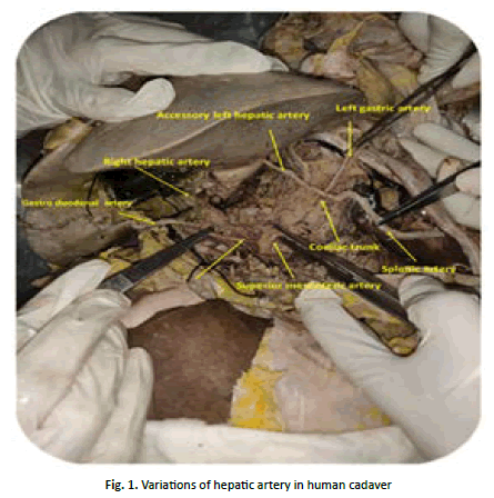
• The common hepatic artery has been arised from superior mesenteric artery and given gastro duodenal artery. Which normally arises from coeliac trunk
• Accessory hepatic artery was arising from coeliac trunk (Figure 1).
Fig. 1. Variations of hepatic artery in human cadaver.
Discussion
The present study correlating with the type IX anatomical variations of the hepatic arterial system were defined according to Michels's, et al., internationally recognized classification and also correlating with the type V, Hiatt's, et al., modification of that system (Table 2) [5].
The present study also correlating with the type I anatomical variations of the hepatic arterial system were found by Geraldo Alberto Sebben, et al. Variations of hepatic artery: Anatomical study on cadavers Geraldo Alberto Sebben, et al. (Table 3) [6,7].
| S.No | Hepatic artery variation | Michels classification | Hiatt classification |
|---|---|---|---|
| 1. | Normal anatomy. | Type I | Type I |
| 2. | Replaced left hepatic artery originating from the left gastric artery. | Type II | Type II |
| 3. | Replaced right hepatic artery originating from the superior mesenteric artery. | Type III | Type III |
| 4. | Co-existence of type II and III. | Type IV | Type IV |
| 5. | Accessory left hepatic artery originating from the left gastric artery. | Type V | Type II |
| 6. | Accessory right hepatic artery originating from the superior mesenteric artery. | Type VI | Type III |
| 7. | Accessory left hepatic artery originating from the left gastric artery and accessory right hepatic artery originating from the superior mesenteric artery. | Type VII | Type IV |
| 8. | Accessory left hepatic artery originating from the left gastric artery and replaced right hepatic artery originating from the superior mesenteric artery. | Type VIII | Type IV |
| 9. | Common hepatic artery originating from the superior mesenteric artery. | Type IX | Type V |
| 10. | Right and left hepatic arteries originating from the left gastric artery. | Type VIII | Type IV |
| 11. | Common hepatic artery directly originating from the aorta. | Type IX | Type V |
Tab. 2. This table shows the anatomical variations of the hepatic arterial system according to michels and hiatt classification.
| S. No | Variation | N | % |
|---|---|---|---|
| 1 | Common hepatic artery originating from the superior mesenteric artery. | 2 | 6.66% |
| 2 | Right gastric artery originating from the right hepatic artery. | 2 | 6.66% |
| 3 | Right gastric artery originating from the superior mesenteric artery. | 1 | 3.33% |
| 4 | Right hepatic artery originating from the superior mesenteric artery. | 3 | 10% |
| 5 | Left hepatic artery originating from the superior mesenteric artery | 1 | 3.33% |
| 6 | Cystic artery originating from the proper hepatic artery bifurcation | 1 | 3.33% |
| 7 | Cystic artery originating from the left hepatic artery | 1 | 3.33% |
| 8 | Right gastric artery originating from the left hepatic artery | 1 | 3.33% |
| 9 | Total | 12 | 40% |
Tab. 3. The table shows the anatomical variations of the hepatic arterial system.
Conclusion
Anatomical variations of the hepatic arteries and coeliac trunk are of considerable importance in liver transplants, laparoscopic surgery, radiological abdominal interventions and penetrating injuries to the abdomen. Knowledge of such variations will play a significant role in avoiding technical difficulties during infusion therapy and chemoembolization of neoplasm in the liver. The frequency of inadvertent or iatrogenic hepatic vascular injury rises in the event of aberrant anatomy and variations. Arterial vascularisation of the gastrointestinal system is provided by anterior branches at three different levels of the abdominal aorta (the coeliac trunk and the superior and inferior mesenteric arteries). Differences arising and branching pattern during several developmental stages in the embryonal process lead to a range of variations in these vascular structures clinicians and radiologist should be aware of such aberrant vascular anatomy so as to reduce the incidence of surgical complication and hepatic cancer. Vascularization from the hepatic artery and its branches is responsible for the vast majority of hepatocellular carcinomas. The principal variants of the hepatic arterial anatomy must be identified on the pre-treatment CT scan or arteriography in order to perform chemoembolization, which requires checking the hepatic arterial anatomy.The extra-hepatic arteries that the tumour recruits may help these tumours develop an abnormal, "parasitic," vascularization. Large peripheral, subcapsular, exophytic tumours, a history of resection, hepatectomy, or repeated chemoembolization are some known risk factors that need to be watched out for. The presence of a vascular pedicle belonging to the tumour itself or a big diameter extra-hepatic artery travelling in the direction of the liver on the pre-treatment CT scan is further proof that the tumour has developed a "parasitic" vascular supply.
References
- Romanes GJ. Cunningham’s manual of practical anatomy. (15th edition). Oxford Medical Publication, Oxford University Press, Oxford. 1986;91-127.
- Michels NA. Newer anatomy of the liver and its variant blood supply and collateral circulation. Am J Surg.1966; 112:337–347.
[Crossref] [Google Scholar] [PubMed]
- Hiatt JR, Gabbay J, Busuttil RW. Surgical anatomy of the hepatic arteries in 1000 cases. AnnSurg.1994; 220:50–52.
[Crossref] [Google Scholar] [PubMed]
- Ugurel MS, Battal B, Bozlar U, Nural MS, Tasar M, et al. Anatomical variations of hepatic arterial system, coeliac trunk and renal arteries: An analysis with multidetector CT angiography. Br J Radiol. 2010; 83:661-667.
[Crossref] [Google Scholar] [PubMed]
- Sebben GA, Rocha SL, Sebben MA, Parussolo Filho PR, Gonçalves BH. Variations of hepatic artery: Anatomical study on cadavers. Rev Col Bras Cir. 2013; 40:221-226.
[Crossref] [Google Scholar] [PubMed]
- Munshi IA, Fusco D, Tashjian D, Kirkwood JR, Polga J, et al. Occlusion of an aberrant right hepatic artery, originating from the superior mesenteric artery, secondary to blunt trauma. J Trauma. 2000; 48:325–326.
[Crossref] [Google Scholar] [PubMed]
- Rela M, McCall JL, Karani J, Heaton ND. Accessory right hepatic artery arising from the left. Transplantation. 1998; 66:792–794.
[Crossref] [Google Scholar] [PubMed]
Copyright:
This is an open access article distributed under the terms of the Creative Commons Attribution License, which permits unrestricted use, distribution, and reproduction in any medium, provided the original work is properly cited.


