Case Report - Onkologia i Radioterapia ( 2026) Volume 20, Issue 9
Penile gangrene in a patient with poorly controlled type 2 diabetes Mellitus: a case report
Donat Cuni, Flatra Fetahu and Xhevdet Cuni*Xhevdet Cuni, Department of Medicine, University of Prishtina, Prishtina, Kosovo, Email: drxhevdetcuni@gmail.com
Received: 05-Aug-2026, Manuscript No. OAR-26-194817; , Pre QC No. OAR-26-194817 (PQ); Editor assigned: 07-Aug-2026, Pre QC No. OAR-26-194817 (PQ); Reviewed: 21-Aug-2026, QC No. OAR-26-194817; Revised: 11-Sep-2026, Manuscript No. OAR-26-194817 (R); Published: 18-Sep-2026
Abstract
Background: Ischemic penile gangrene is an uncommon manifestation of advanced vascular and metabolic disease. Because genital tissue necrosis can mimic or coexist with infection, malignancy, treatment-related injury, or other destructive processes, early clinical characterization is important. In this report, I will discuss the disease called distal penile gangrene in a person suffering from severe diabetes mellitus and existing ischemia of the lower extremities.
Case presentation: 67-year-old male with poorly controlled type 2 diabetes for more than 10 years was admitted to the hospital with the problem of gradual blackening of the penis glans. Patient suffers from severe lesion of vessels of the lower extremities and amputation of the right lower limb below the knee. In examination, dry gangrene was revealed on the surface of small focus without pus discharge, without affection of the testicles and lymphadenitis of inguinal lymph nodes. The level of HbA1c is 13%. After consultation of multidisciplinary team and preparation of the patient in optimal condition, necrotic lesion of glans penis was surgically removed. Opening of urinary tract was restored with insertion of catheter in urethra. Recovery from the surgery was adequate to be classified as positive, and there were no complications in the early postoperative period. Upon completion of rehabilitation therapy, the patient was transferred to a special unit with prolonged care.
Conclusion: Distal penile gangrene must be assessed for the presence of underlying systemic disorders of the vascular system and excluded any infections and other factors causing necrosis. Tissue-preserving surgery may restore urinary function in the presence of necrotic tissue in certain locations and with well-defined borders. Differentiation of ischemic necrosis from non-ischemic lesions of the penis is very important for further diagnostic approach in case of malignancy of the penis.
Keywords
Penile gangrene; Penile ischemia; Penile necrosis; Diabetes mellitus; Peripheral vascular disease; Tissue necrosis; Partial penectomy; Case report
Introduction
Penile gangrene is a rare disease characterized by a necrosis of genital tissues caused by an extremely decreased perfusion in this area. In normal circumstances, the penis receives a good blood supply due to numerous arteries; thus, the tissue damage or death results from rather serious disorders such as insufficient blood flow, metabolic problems, infections, etc.
Long-term suffering from diabetes is associated with a number of complications, including problems with blood vessel lining, accelerated formation of arterial plaques, microangiopathy, neuropathy, and a slower wound healing process [1]. The presence of both diabetes and severe peripheral vascular disease will result in the overall decrease of the blood supply to the tissues making them more vulnerable due to the lack of it [2].
Thus, the main distinguishing feature of the initial stage of dry ischemic gangrene and infection necrosis should be highlighted. Ischemic dry gangrene appears as sharply defined black or dried tissue without any signs of pus or discharge. The wet gangrene and infections associated with tissue death will be complicated with an additional infection of the affected area and will progress rather fast [3]. Among the other potential diseases one should mention calciphylaxis, ischemia due to trauma, severe inflammation or infection.
For oncology-oriented clinical assessment, destructive penile lesions require careful correlation of morphology, history, vascular status, infection findings, and when clinically indicated histopathology [4]. In patients with a history of cancer therapy, treatment-related tissue injury would also need to be distinguished from recurrent or progressive disease; however, no prior oncologic treatment or radiotherapy was documented in the present case.
We report a rare case of distal penile gangrene in a 67-year-old man with markedly uncontrolled diabetes mellitus and severe peripheral vascular disease [5-6]. The case emphasizes clinical differentiation of ischemic tissue necrosis and the role of tissue-preserving surgical management.
Case Presentation
A 67-year-old man was referred to the emergency center because of progressive dark discoloration of the distal penis. His medical history included type 2 diabetes mellitus diagnosed more than 10 years earlier and severe psychiatric disease treated with antipsychotic medication [7]. Communication was difficult at presentation, and psychiatric consultation was obtained.
The patient had poor adherence to prescribed oral hypoglycemic therapy [8]. Seven years before the current presentation, he had undergone a right below-knee amputation because of severe peripheral vascular disease.
The principal presenting finding was progressive black discoloration of the glans and distal penile tissue [9-10]. Physical examination demonstrated superficial dry gangrene (Figure 1). There was no reported purulent discharge, the testes were clinically normal, and no inguinal lymphadenopathy was detected.
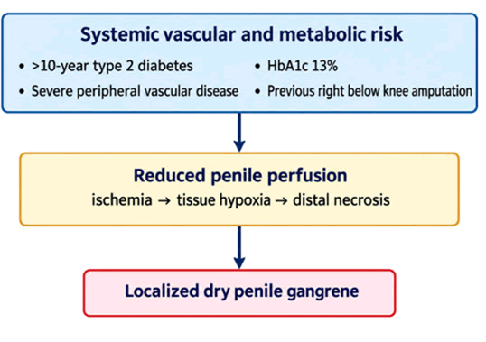
Fig. 1. Schematic pathophysiologic relationship between severe diabetes, peripheral vascular disease, reduced penile perfusion, and distal dry gangrene. The diagram summarizes mechanisms documented or discussed in the case; it is not a photograph of the patient.
The available clinical material documents an HbA1c concentration of 13%, consistent with severe chronic hyperglycemia [11]. Other laboratory results were not available in the source clinical material and therefore are not reported here.
The lesion was considered clinically compatible with localized dry penile gangrene because of its appearance and the absence of documented purulent discharge, testicular involvement, or clinical evidence of ascending perineal infection [12]. Given the patient's severe peripheral vascular disease and poorly controlled diabetes, ischemic penile necrosis was considered the most likely mechanism.
After multidisciplinary evaluation and preoperative preparation, the patient underwent surgery under general endotracheal anesthesia. The necrotic distal penile tissue was resected while viable tissue was preserved. The urethral opening was reconstructed and a transurethral catheter was inserted (Figures 2-4).
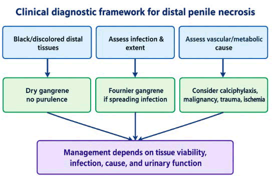
Fig. 2. Schematic diagnostic framework for a black or necrotic distal penile lesion, emphasizing assessment for infection, vascular/metabolic causes, calciphylaxis, malignancy, trauma, and other etiologies. The branches are conceptual and should be interpreted with clinical findings.
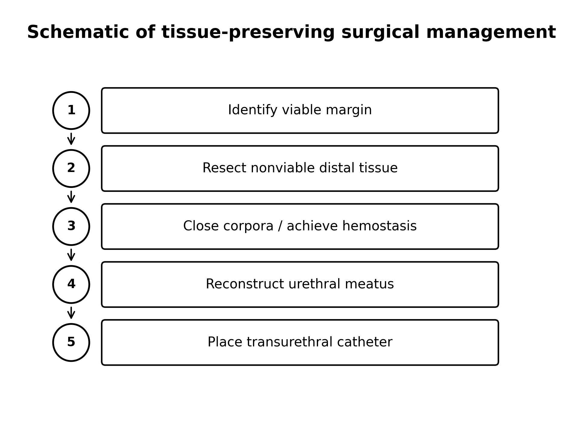
Fig. 3. Schematic sequence of the tissue-preserving surgical approach described in the case: Identification of viable tissue, resection of nonviable tissue, closure/ hemostasis, urethral reconstruction, and catheter placement.
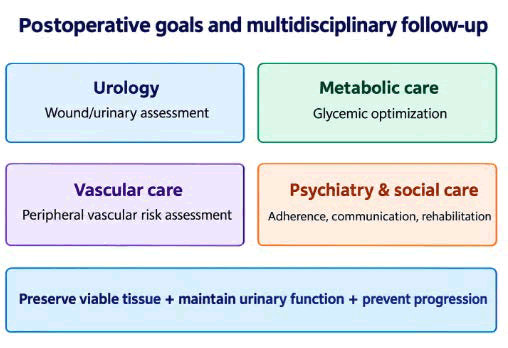
Fig. 4. Schematic multidisciplinary follow-up framework emphasizing urological care, metabolic optimization, vascular risk assessment, psychiatric/social support, urinary function, and prevention of progression.
The postoperative course was clinically favorable, with no immediate major surgical complication reported. After rehabilitation and stabilization, the patient was transferred to a specialized long-term care facility.
Surgical technique
Following induction of general endotracheal anesthesia, the extent of devitalized tissue was assessed. The necrotic distal segment was resected until viable tissue was identified. The corpora cavernosa were closed to achieve hemostasis and reconstruct the distal anatomy. The urethra was mobilized and reconstructed to establish an adequate urethral meatus, and a transurethral catheter was placed to facilitate urinary drainage during the early postoperative period.
The operative objective was removal of nonviable tissue with maximal preservation of viable penile length and maintenance of urinary function. The surgical sequence is presented schematically in Table 1.
| Clinical event | Description |
| >10 years before presentation | Diagnosis of type 2 diabetes mellitus |
| 7 years before presentation | Right below-knee amputation for severe peripheral vascular disease |
| Before current admission | Poor adherence to oral antidiabetic therapy; chronic psychiatric disease treated with antipsychotic medication |
| Current presentation | Progressive black discoloration of the distal penis |
| Emergency evaluation | Psychiatric consultation and urological assessment |
| Laboratory assessment | HbA1c 13% |
| Preoperative period | Multidisciplinary assessment and preparation for surgery |
| Surgical treatment | Resection of necrotic penile tissue and urethral reconstruction |
| Postoperative period | Favorable clinical recovery and rehabilitation |
| Discharge/transfer | Transfer to specialized long-term psychiatric care |
Tab. 1. Clinical timeline of disease progression, evaluation, surgical management, and postoperative course.
Diagnostic and oncology-relevant considerations
Although the present case is not a radiotherapy-related malignancy case, penile tissue necrosis has an important differential-diagnostic relationship with oncology. A destructive penile lesion may be caused by ischemia, infection, malignancy, treatment-related injury, or combinations of these processes.
Results and Discussion
The present case illustrates distal penile gangrene occurring in the setting of severe metabolic dysregulation and established peripheral vascular disease. The unusual location of the ischemic lesion is clinically important because penile necrosis is uncommon despite the organ's extensive arterial collateralization.
The patient's HbA1c of 13% indicates marked chronic hyperglycemia. Diabetes may impair tissue viability through several interacting mechanisms, including endothelial dysfunction, atherosclerotic disease, microvascular injury, neuropathy, and impaired wound healing. The previous below-knee amputation provides additional clinical evidence of substantial peripheral vascular disease in this patient.
The clinical phenotype was consistent with dry rather than wet gangrene. The lesion was described as superficial and black, without purulent discharge, testicular involvement, or documented perineal extension. This distinction is clinically important because necrotizing infection, including Fournier's gangrene, requires urgent recognition and treatment and can extend beyond the initially visible area.
Calciphylaxis has also been mentioned as one of the causes for penile tissue necrosis, particularly in individuals with advanced chronic kidney disease and diabetes. In the clinical data presented in this report, no evidence of chronic kidney disease, dialysis, high serum calcium level, or histopathologic confirmation of calciphylaxis is found. Therefore, it is not appropriate to suggest calciphylaxis as a cause of this particular condition.
It should also be remembered that penile cancer can be one of the possible causes of persistent, painful, hard and/or abnormalappearing lesions of the penis. When working with a cancer patient, special attention needs to be paid to the history of prior malignancies and its treatment, imaging studies, pathology report. Prior local radiotherapy in a patient can be considered as one of the potential causes. In the current case, there is no data on radiation history of this patient.
There is also another possible cause of death of penile tissues known as calciphylaxis; this disease is common in patients with serious kidney conditions and diabetes. The clinical presentation provided in the report, however, does not indicate symptoms such as chronic renal failure, dialysis treatment, increased level of calcium and biopsy findings of calciphylaxis. It is, thus, not possible to prove that this is the cause of death of penile tissue in this case.
The patient may have developed cancer of the penis which would explain the presence of persistent, ulcerated, hard and unusual looking lesion on his penis. In an oncological setting, a thorough evaluation of the past history of cancer in the patient as well as his imaging results and treatment history is very vital. When the patient has received local radiotherapy in the past, one can consider the radiation treatment as possible cause of tissue death. There is, however, no history of cancer and radiotherapy in this case.
Conclusion
Localized penile gangrene is one of the rarest manifestations of vascular complications and metabolic diseases. Among those patients who suffer from poorly controlled diabetes for many years and have concomitant circulatory disorders in legs, any changes of color on the head or foreskin of the penis should be investigated concerning such possible causes as ischemia, infectious processes, and other factors leading to necrosis.
The approach of tissue-preserving resection combined with urethral reconstruction provides excellent management of urinary function in case of localized necrosis with possibility of preservation of healthy tissue. Multiple medical issues require involvement of various specialists, especially, in presence of several concomitant diseases such as metabolic disorders, vascular pathology, psychiatric disorders, and social dysfunction.
When analyzing the cases of cancer, the principal task is to distinguish ischemic necrosis from possible tumors, infectious, calcific, traumatic, or iatrogenic origin. There is no history of malignant disorders or radiotherapy of the patient, that is why the report discusses possible malignant diseases of the patient instead of data about radiotherapy.
Funding
No specific funding was received for this case report.
Conflict of Interest
The authors declare that they have no conflicts of interest related to this manuscript.
Author Contributions
All authors contributed to the clinical management, interpretation of the case, manuscript preparation, critical revision, and approval of the final manuscript.
Patient Consent
A case report involving identifiable clinical information and/or clinical images should be submitted only after obtaining appropriate written informed consent in accordance with the journal's requirements. The source manuscript provided for revision does not contain a consent statement; this should be completed by the authors before submission.
Ethics Statement
The appropriate institutional requirements for publication of a case report should be confirmed by the authors and stated according to the journal's instructions.
References
- Bour J, Steinhardt G. Penile necrosis in patients with diabetes mellitus and end stage renal disease. J Urol. 1984; 132:560-562.
[Crossref] [Google Scholar] [PubMed]
- Nomura T, Sakamoto F. A case of penile necrosis in a patient with diabetes mellitus. Nihon Hinyokika Gakkai Zasshi. 1992; 83:1138-1141.
[Crossref] [Google Scholar] [PubMed]
- Dunev VR, Kolev NH, Genov PP. A rare clinical case of penile gangrene in a patient with chronic renal failure and diabetes mellitus. Urol Case Rep. 2019; 23:108-109.
[Crossref] [Google Scholar] [PubMed]
- López Pacios JC, Sánchez Merino JM, Piñeiro Fernández MC, Parra Muntaner L, Gómez Cisneros SC, et al. Necrosis of the penis in a diabetic patient: Presentation of a case. Arch Esp Urol. 2003; 56:434-436.
[Google Scholar] [PubMed]
- Rajbabu K, Browning AJ, Parsons KF. Gangrene of the penis in a diabetic male with multiple amputations and follow up. J Med Case Rep. 2009; 3:9321.
[Crossref] [Google Scholar] [PubMed]
- Aladren M. Surgical management of ischemic penile gangrene in diabetics with end stage atherosclerosis. J Urol. 1996; 155:926-929.
[Google Scholar] [PubMed]
- Dogru T. Penile gangrene: A rare complication of diabetes mellitus. Pract Diabetes Int. 2006; 23:273-275.
- Al Hadidi S, Haddad RA, Abed F, Bachuwa G. Penile gangrene as a sign of uncontrolled diabetes mellitus. BMJ Case Rep. 2016.
[Crossref] [Google Scholar] [PubMed]
- Grandez-Urbina JA. Case Report: Penile necrosis associated to paraphimosis with calciphylaxis due to terminal chronic kidney disease. F1000Res. 2019; 8:139.
[Crossref] [Google Scholar] [PubMed]
- Chiang IN, Chang SJ, Kuo YC, Liu SP, Yu HJ, et al. Management of ischemic penile gangrene: Prompt partial penectomy and other treatment options. J Sex Med. 2008; 5:2725-2733.
[Crossref] [Google Scholar] [PubMed]
- Gagnier JJ, Kienle G, Altman DG, Moher D, Sox H, et al. The CARE guidelines: Consensus-based clinical case reporting guideline development. BMJ Case Rep. 2013; 2013:bcr2013201554.
[Crossref] [Google Scholar] [PubMed]
- Riley DS, Barber MS, Kienle GS, Aronson JK, von Schoen-Angerer T, et al. CARE guidelines for case reports: Explanation and elaboration document. J Clin Epidemiol. 2017; 89:218-235.
[Crossref] [Google Scholar] [PubMed]