Research Article - Onkologia i Radioterapia ( 2026) Volume 20, Issue 8
Neurological Manifestations, Treatment Outcomes, and Survival Determinants in Patients with Gliomas Undergoing Postoperative Radiotherapy: A Retrospective Cohort Study from Central Asia
Nargiza Sunnatillayevna Karimova1,2, Daniyar Anarbayevich Nishanov2, Khilola Bakhtiyarovna Abdakhatova1, Yashnar Mamasaliyevna Mamadaliyeva3, Anvar Pardabayevich Tuychiyev1,2, Khojiakbar Khamid ugli Khamidov2 and Mukhayo Ravshanovna Khayitboeva4*2Tashkent State Medical University, Tashkent, Uzbekistan
3Center for the Development of Professional Qualification of Medical Workers, Tashkent, Uzbekistan
4Kimyo International University in Tashkent, Tashkent, Uzbekistan
Mukhayo Ravshanovna Khayitboeva, Kimyo International University in Tashkent, Tashkent, Uzbekistan, , khayitboyeva@list.ru
Received: 01-Aug-2026, Manuscript No. OAR-26-194568; , Pre QC No. OAR-26-194568; Editor assigned: 04-Aug-2026, Pre QC No. OAR-26-194568; Reviewed: 18-Aug-2026, QC No. OAR-26-194568; Revised: 24-Aug-2026, Manuscript No. OAR-26-194568; Published: 29-Aug-2026
Abstract
Background: Malignant gliomas are among the most aggressive primary brain tumors and are associated with poor prognosis despite multimodal treatment. Data regarding neurological complications and prognostic factors in patients with malignant gliomas from Central Asian populations remain limited. This study aimed to evaluate neurological manifestations, treatment-related toxicity, recurrence patterns, and survival-related prognostic factors in patients with malignant gliomas undergoing postoperative radiotherapy. Methods: This retrospective cohort study included 174 consecutive patients with histologically confirmed malignant gliomas treated with surgery followed by radiotherapy between 2018 and 2023. Clinical, radiological, histopathological, and immunohistochemical data were analyzed. Overall survival was estimated using the Kaplan–Meier method, and independent prognostic factors were identified using multivariable Cox proportional hazards regression. Results: Headache (91.4%), general weakness (74.7%), and seizures (47.1%) were the most common neurological manifestations. Three-dimensional conformal radiotherapy was the most frequently used treatment modality (58.0%). Acute post-treatment findings included cerebral edema (83.3%), alopecia (92.5%), and radiation dermatitis (79.9%), whereas persistent headache (87.9%), epilepsy (52.3%), memory impairment (54.0%), and cognitive dysfunction (50.0%) predominated among late follow-up findings. During follow-up, 84 deaths were recorded, and the median overall survival for the entire cohort was 48 months. Kaplan–Meier analysis demonstrated significantly shorter survival in patients with WHO grade IV tumors. Multivariable Cox regression identified higher WHO grade as the only independent predictor of worse overall survival (HR=1.72, 95% CI 1.06–2.78, p=0.028), whereas age, sex, Karnofsky Performance Status, extent of tumor reduction, and radiotherapy technique were not independently associated with survival. Conclusions: Neurological manifestations and long-term outcomes in patients with malignant gliomas were associated with clinical, anatomical, pathological, and treatment-related characteristics. Histological grade represented the principal prognostic factor associated with overall survival in the present cohort, while a substantial burden of acute and delayed neurological findings was documented during follow-up after radiotherapy. Continuous neurological monitoring and multidisciplinary management remain important components of patient care.
Keywords
Malignant glioma; Glioblastoma; Postoperative radiotherapy; Neurological complications; Survival analysis; Prognostic factors; Cox regression
INTRODUCTION
Gliomas comprise a heterogeneous group of primary central nervous system tumors with substantial variation in histopathological characteristics, molecular profiles, clinical presentation, treatment response, and survival. Despite advances in neurosurgery, radiotherapy, systemic therapy, and neuroimaging, patients with high-grade gliomas continue to experience considerable morbidity and mortality. Glioblastoma, in particular, remains associated with an unfavorable prognosis despite multimodal treatment [6,10,11,15].
Neurological manifestations are common at the time of glioma diagnosis and are influenced by tumor location, size, mass effect, peritumoral edema, and involvement of functionally important brain structures. Headache, seizures, cognitive impairment, focal motor deficits, and disturbances of coordination may substantially affect patients' functional status and quality of life. Importantly, these manifestations may be present before the initiation of oncological treatment and should therefore be distinguished from adverse events developing during or after treatment [1,7,10]. Characterizing baseline neurological presentation according to anatomical tumor localization may provide clinically relevant information for patient assessment and follow-up.
Postoperative radiotherapy remains an important component of multimodal management for patients with gliomas who meet appropriate clinical and pathological indications. Depending on tumor characteristics and clinical circumstances, radiotherapy may be administered with concurrent systemic treatment, most commonly temozolomide in selected high-grade gliomas [8,11]. In routine clinical practice, however, treatment selection is influenced by tumor histology, anatomical location, performance status, age, comorbidities, treatment availability, and patient preferences. Consequently, real-world cohorts may demonstrate substantial variation in radiotherapy techniques and systemic treatment. These differences are important when evaluating treatment-related adverse events and survival outcomes because treatment selection is not necessarily independent of underlying disease characteristics.
The biological heterogeneity of gliomas has become increasingly important with the introduction of integrated molecular classification. The 2021 World Health Organization Classification of Tumors of the Central Nervous System emphasizes the integration of histopathological and molecular characteristics, including alterations such as IDH mutation and 1p/19q codeletion, for the classification of diffuse gliomas [3]. Other molecular and immunohistochemical characteristics, including MGMT promoter methylation, Ki-67 proliferative activity, and selected molecular or protein-expression markers, may also provide prognostic information [9]. However, retrospective cohorts assembled over several years may contain incomplete molecular characterization, particularly when diagnostic practices and availability of molecular testing have changed over time. The extent and pattern of missing molecular information therefore need to be considered when interpreting prognostic analyses.
Although neurological presentation, treatment-related adverse events, recurrence, and survival have each been investigated in glioma populations, integrated real-world data from Central Asian patients remain limited. Previous studies from Uzbekistan have primarily addressed neuroimaging characteristics and radiotherapy planning, including the application of multiparametric magnetic resonance imaging and imaging-based approaches to clinical target-volume delineation [2,4]. Less is known about the distribution of baseline neurological manifestations according to tumor localization, patterns of postoperative radiotherapy and concurrent systemic treatment, recurrence, and survival in routine clinical practice in this region.
The present study therefore aimed to characterize baseline neurological manifestations, postoperative treatment patterns, treatment-related adverse events, recurrence patterns, and survival outcomes in a retrospective cohort of patients with histologically diagnosed gliomas undergoing postoperative radiotherapy in Uzbekistan. A secondary objective was to identify clinical, radiological, pathological, and treatment-related factors associated with overall survival while taking into account the limitations of incomplete molecular characterization.
MATERIALS AND METHODS
Study design and patient population
This retrospective cohort study included 174 consecutive patients with histologically confirmed gliomas who underwent surgical treatment followed by postoperative radiotherapy at the Republican Specialized Scientific-Practical Medical Center of Oncology and Radiology and its regional branches between January 2018 and December 2023.
Patients were eligible if they had: (1) histologically confirmed glioma; (2) available preoperative and postoperative magnetic resonance imaging (MRI); (3) postoperative radiotherapy as part of their treatment; and (4) available clinical and follow-up information. Patients without histological verification or with insufficient clinical or follow-up information were excluded.
Histopathological diagnoses and tumor grades were recorded according to the institutional pathology reports. Molecular and immunohistochemical findings were recorded when available. Because molecular testing was not performed in all patients, these data were considered incompletely available and were analyzed accordingly rather than being assumed for patients without testing.
Clinical and radiological assessment
Demographic, clinical, radiological, surgical, pathological, and treatment-related data were retrospectively extracted from medical records.
The collected variables included age, sex, Karnofsky Performance Status (KPS), neurological manifestations at presentation, anatomical tumor location, primary tumor size, extent of surgical resection, postoperative residual tumor, histological grade, available molecular and immunohistochemical markers, radiotherapy technique, radiation dose, systemic treatment, follow-up MRI findings, and survival outcomes.
Neurological manifestations documented before initiation of postoperative radiotherapy included headache, dizziness, seizures, nausea, loss of consciousness, memory impairment, limb weakness, and general weakness. Baseline neurological manifestations were analyzed separately from symptoms documented during follow-up and were not automatically considered radiotherapy-related adverse events.
Primary tumor size was assessed on preoperative imaging according to the measurements documented in the medical records. Postoperative MRI was used to assess the presence and size of residual tumor.
Histopathological, molecular, and immunohistochemical assessment
Histological grade was recorded as documented in the institutional pathology reports. Available immunohistochemical and molecular investigations included Ki-67 proliferative activity, p53 expression, vascular endothelial growth factor (VEGF), IDH1/IDH2 mutation status, MGMT promoter methylation, and 1p/19q codeletion.
The availability of each molecular or immunohistochemical marker was recorded separately. Because these investigations were not available for the entire cohort, incomplete molecular characterization was considered a limitation of the retrospective dataset. No molecular status was assigned to patients without corresponding laboratory or pathology results.
Surgery and assessment of residual disease
The extent of surgical intervention was categorized as total, subtotal, or partial resection according to the postoperative clinical and radiological documentation.
Postoperative MRI was used to determine whether residual tumor was present and, when documented, to categorize residual tumor size. The presence of residual disease and the recorded extent of resection were analyzed as separate variables.
Radiotherapy and systemic treatment
Postoperative radiotherapy was delivered using three-dimensional conformal radiotherapy (3D-CRT), intensity-modulated radiotherapy (IMRT), or volumetric modulated arc therapy (VMAT). Treatment planning was performed using computed tomography with magnetic resonance imaging correlation. Total radiation doses ranged from 50 to 60 Gy and were prescribed according to tumor characteristics, target volume, histological grade, and clinical treatment decisions.
Systemic treatment was recorded from the medical records. Temozolomide was the principal systemic agent used in the cohort. Other documented systemic agents included irinotecan, lomustine, and bevacizumab. Patients who did not receive concurrent systemic treatment were also identified. Reasons documented for omission of systemic treatment included less aggressive histology, poor performance status, severe comorbidities, contraindications, and patient refusal.
Because this was a retrospective observational study, radiotherapy technique and systemic treatment were selected according to clinical and anatomical characteristics. Therefore, associations between treatment variables and outcomes were interpreted as observational associations and not as evidence of causal treatment effects.
Follow-up and outcome assessment
Patients underwent clinical and radiological follow-up after treatment. Follow-up MRI examinations were reviewed for documented residual disease, progression, recurrence, cystic changes, hemorrhagic changes, and other intracranial abnormalities.
Overall survival (OS) was defined as the interval from the date of histological diagnosis to death from any cause or the date of the last available follow-up for patients who remained alive.
Progression-free survival (PFS) was defined as the interval from the date of histological diagnosis to the first documented disease progression or death from any cause, whichever occurred first. Patients without documented progression or death were censored at the date of the last available follow-up.
Because of the retrospective nature of the study, radiological findings were analyzed according to the information available in the medical records. When standardized Response Assessment in Neuro-Oncology (RANO) criteria were not explicitly documented, follow-up MRI findings were not retrospectively classified as definite progression, pseudoprogression, or radiation necrosis.
Assessment of post-treatment adverse events
Clinical events documented during and after radiotherapy were extracted from medical records and categorized according to the timing documented in the records.
Documented acute events included gastrointestinal symptoms, hematological abnormalities, radiation-related skin reactions, alopecia, and neurological symptoms occurring during or shortly after radiotherapy. Late follow-up findings included persistent headache, memory impairment, seizures or epilepsy, cognitive dysfunction, and paresis.
Baseline neurological manifestations documented before radiotherapy were analyzed separately from post-treatment events. Because the retrospective records did not provide standardized prospective toxicity grading, systematic baseline neuropsychological testing, or uniform assessment of alternative causes of neurological symptoms, post-treatment neurological findings were not assumed to be definitively caused by radiotherapy.
Study endpoints
The primary endpoint was overall survival
Secondary endpoints included neurological manifestations at presentation, postoperative residual disease, radiotherapy and systemic treatment patterns, documented post-treatment adverse events, disease progression and recurrence, progression-free survival, and factors associated with overall survival.
Statistical analysis
Statistical analyses were performed using IBM SPSS Statistics version 27.0 (IBM Corp., Armonk, NY, USA). Continuous variables were summarized as mean ± standard deviation or median with interquartile range, as appropriate. Categorical variables were presented as frequencies and percentages. Associations between categorical variables were assessed using the chi-square test or Fisher's exact test, as appropriate. Continuous variables were compared using the Student's t-test or Mann–Whitney U test according to data distribution. Overall survival and progression-free survival were estimated using the Kaplan–Meier method. Survival distributions between groups were compared using the log-rank test. Cox proportional hazards regression was used to evaluate factors associated with overall survival. Candidate variables included age, sex, KPS, histological grade, extent of surgical resection/tumor reduction, and radiotherapy technique. Hazard ratios (HRs) with 95% confidence intervals (CIs) were reported. The proportional hazards assumption was assessed before interpretation of the multivariable Cox model. Variables with substantial missingness were reported as missing and were not assigned values based on assumptions. Molecular and immunohistochemical variables were interpreted in the context of their limited availability. Because treatment allocation was not randomized and radiotherapy technique was associated with anatomical tumor characteristics, treatment-related associations were interpreted with consideration of potential confounding by indication. All statistical tests were two-sided, and a p-value <0.05 was considered statistically significant.
RESULTS
Baseline characteristics of the study population
A total of 174 patients with histologically confirmed malignant gliomas were included in the study. Baseline demographic, clinical, radiological, and histopathological characteristics are summarized in Table 1.
The study population consisted predominantly of male patients (55.7%), whereas females accounted for 44.3% of the cohort. Patients aged 20–39 years represented the largest age group (46.5%). At admission, most patients presented with moderate clinical condition (46.6%), while severe or very severe status was observed in only 4.6% of cases.
Subtotal tumor resection represented the most common surgical approach and was performed in 75.3% of patients. Total resection was achieved in 17.8%, whereas partial resection accounted for 6.9% of cases.
Histopathological examination demonstrated a predominance of high-grade tumors. WHO grade III gliomas accounted for 73.0% of all cases, while grade IV tumors represented 17.8%. Grade II tumors were relatively uncommon and were observed in 9.2% of patients.
Overall, the study cohort was characterized by a predominance of high-grade gliomas, extensive residual tumor burden, and subtotal surgical resection, reflecting the aggressive nature of the disease and the complexity of treatment [Table 1].
| Characteristic | Category | n (%) |
|---|---|---|
| Sex | Male | 97 (55.7) |
| Female | 77 (44.3) | |
| Age group (years) | 0–9 | 3 (1.7) |
| 10–19 | 19 (10.9) | |
| 20–29 | 40 (23.0) | |
| 30–39 | 41 (23.6) | |
| 40–49 | 34 (19.5) | |
| 50–59 | 25 (14.4) | |
| 60–69 | 12 (6.9) | |
| General condition at admission | Satisfactory | 53 (30.5) |
| Relatively satisfactory | 32 (18.4) | |
| Moderate severity | 81 (46.6) | |
| Severe | 7 (4.0) | |
| Very severe | 1 (0.6) | |
| Extent of surgical resection | Total resection | 31 (17.8) |
| Subtotal resection | 131 (75.3) | |
| Partial resection | 12 (6.9) | |
| Primary tumor size | <2 cm | 6 (3.4) |
| 2–4 cm | 43 (24.7) | |
| 4–5 cm | 45 (25.9) | |
| >5 cm | 80 (46.0) | |
| Residual tumor size on postoperative MRI | 1–2 cm | 51 (32.7) |
| 3–4 cm | 74 (47.4) | |
| 5–6 cm | 15 (9.6) | |
| 7–8 cm | 15 (9.6) | |
| 9–10 cm | 1 (0.6) | |
| No residual tumor | 18 (10.3) | |
| Histological grade | Grade II (G2) | 16 (9.2) |
| Grade III (G3) | 127 (73.0) | |
| Grade IV (G4) | 31 (17.8) |
Table 1: Baseline demographic and clinicopathological characteristics of patients with malignant gliomas (n=174).
Neurological manifestations according to tumor localization
Headache was the most common presenting symptom and was observed in 91.4% of patients. General weakness was reported by 74.7% of patients, whereas dizziness and seizures were documented in 47.1% of cases. Nausea occurred in 37.9% of patients, while memory impairment and loss of consciousness were less frequent manifestations.
Neurological symptoms varied significantly according to anatomical tumor localization (χ²=55.667, p=0.0017). Frontal and temporal lobe tumors demonstrated the highest neurological symptom burden, whereas cerebellar and brainstem lesions were more frequently associated with coordination disorders and balance disturbances. The distribution of neurological symptoms according to anatomical tumor location is shown in [Figure 1].
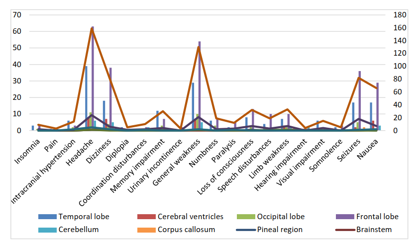
Figure 1: Distribution of neurological symptoms according to anatomical tumor location.
These findings indicate that clinical presentation in malignant gliomas is strongly influenced by tumor topography.
Radiotherapy characteristics
Radiotherapy techniques varied according to anatomical tumor location (χ²=20.869, p=0.0133). Overall, three-dimensional conformal radiotherapy (3D-CRT) represented the predominant treatment modality and was administered in 58.0% of patients. Intensity-modulated radiotherapy (IMRT) and volumetric modulated arc therapy (VMAT) were used in 35.6% and 6.3% of patients, respectively.
Advanced conformal techniques were more frequently employed for tumors located in anatomically complex regions requiring improved dose conformity and sparing of adjacent critical structures [Table 2].
| Tumor location | 3D-CRT | IMRT | VMAT | Total |
|---|---|---|---|---|
| Frontal lobe | 43 (61.4) | 24 (34.3) | 3 (4.3) | 70 (40.2) |
| Cerebellum | 4 (66.7) | 2 (33.3) | 0 | 6 (3.4) |
| Cerebellar vermis | 1 (20.0) | 2 (40.0) | 2 (40.0) | 5 (2.9) |
| Pineal region | 1 (100.0) | 0 | 0 | 1 (0.6) |
| Thalamic region | 1 (100.0) | 0 | 0 | 1 (0.6) |
| Temporal lobe | 23 (57.5) | 15 (37.5) | 2 (5.0) | 40 (23.0) |
| Parietal lobe | 18 (66.7) | 6 (22.2) | 3 (11.1) | 27 (15.5) |
| Occipital lobe | 5 (38.5) | 8 (61.5) | 0 | 13 (7.5) |
| Corpus callosum | 1 (50.0) | 1 (50.0) | 0 | 2 (1.1) |
| Cerebral ventricles | 4 (44.4) | 4 (44.4) | 1 (11.1) | 9 (5.2) |
| Total | 101 (58.0) | 62 (35.6) | 11 (6.3) | 174 (100) |
Statistical analysis: χ² = 20.869; p = 0.0133.
Table 2: Distribution of Radiotherapy Techniques According to Tumor Location n (%)
Treatment-related toxicity
Acute toxicity was mainly represented by cerebral edema accompanied by headache, nausea, and vomiting (83.3%), alopecia (92.5%), and radiation dermatitis (79.9%). Seizure episodes developed in 43.1% of patients. Hematological toxicity was uncommon and occurred in only 2.9% of cases.
During long-term follow-up, neurological sequelae became the dominant form of late toxicity. Persistent headache was observed in 87.9% of patients, memory impairment in 54.0%, epilepsy in 52.3%, and cognitive dysfunction in 50.0%. Motor deficits in the form of limb paresis were documented in 5.2% of patients.
These findings emphasize the importance of prolonged neurological monitoring and rehabilitation in patients undergoing radiotherapy for malignant gliomas.
Immunohistochemical characteristics
Immunohistochemical evaluation was available only in a subset of patients. IDH status assessment demonstrated positive expression in 24 patients (13.8%), whereas negative expression was observed in 3 patients (1.7%). In the majority of patients, immunohistochemical examination was not performed (147 patients, 84.5%).
Assessment of proliferative activity using the Ki-67 labeling index revealed high expression (≥30%) in 33 patients (19.0%), intermediate expression (15–30%) in 16 patients (9.2%), and low or negative expression in 4 patients (2.3%). Ki-67 analysis was unavailable in 121 patients (69.5%).
These findings demonstrate considerable biological heterogeneity among malignant gliomas. However, the large proportion of patients without immunohistochemical characterization limited the possibility of incorporating molecular variables into multivariable survival analysis. The distribution of immunohistochemical characteristics is presented in [Table 3].
| Characteristic | Category | n (%) |
|---|---|---|
| IDH status | Positive | 24 (13.8) |
| Negative | 3 (1.7) | |
| Not performed | 147 (84.5) | |
| Ki-67 expression | High (≥30%) | 33 (19.0) |
| Intermediate (15–30%) | 16 (9.2) | |
| Low/negative | 4 (2.3) | |
| Not performed | 121 (69.5) |
Table 3: Immunohistochemical characteristics of patients with malignant gliomas (n=174).
MRI findings during follow-up after surgery and chemoradiotherapy
Follow-up magnetic resonance imaging demonstrated heterogeneous postoperative and post-treatment changes in patients with malignant gliomas. Residual tumor tissue and hemorrhagic changes represented the most frequent radiological findings. Disease progression and tumor recurrence were observed in a subset of patients, whereas cyst formation and metastatic lesions were less common.
These findings illustrate the heterogeneous biological behavior of malignant gliomas and emphasize the importance of serial magnetic resonance imaging for long-term surveillance and assessment of treatment response. Detailed MRI findings observed during follow-up are summarized in [Table 4].
| MRI finding | n (%) |
|---|---|
| Hemorrhagic changes | 136 (78.2) |
| Residual tumor tissue | 112 (64.4) |
| Disease progression | 27 (15.5) |
| Tumor recurrence | 8 (4.6) |
| Cyst formation | 74 (42.5) |
| Metastatic lesions | 4 (2.3) |
| Missing data | 2 (1.1) |
Table 4: MRI findings during follow-up after surgery and chemoradiotherapy (n=174).
Survival analysis
Overall survival was evaluated using the Kaplan–Meier method. During follow-up, 84 deaths were recorded. The median overall survival for the entire cohort was 48 months.
Survival curves demonstrated a significant association between histological grade and patient prognosis. Patients with WHO Grade IV gliomas exhibited substantially shorter overall survival compared with patients with Grade II and Grade III tumors [Figure 2]. Loonkologia-Kaplan-20-8-0012-g002g-rank analysis demonstrated statistically significant differences between the survival curves (p < 0.05).
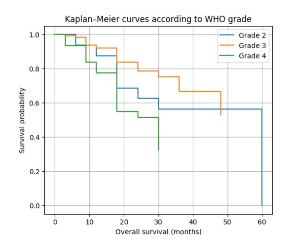
Figure 2: Kaplan–Meier curves according to WHO grade.
Kaplan–Meier analysis according to Karnofsky Performance Status (KPS) showed a tendency toward improved survival among patients with better functional status [Figure 3]. Similarly, patients who achieved greater tumor reduction after surgery demonstrated longer survival compared with those with less extensive resection [Figure 4].
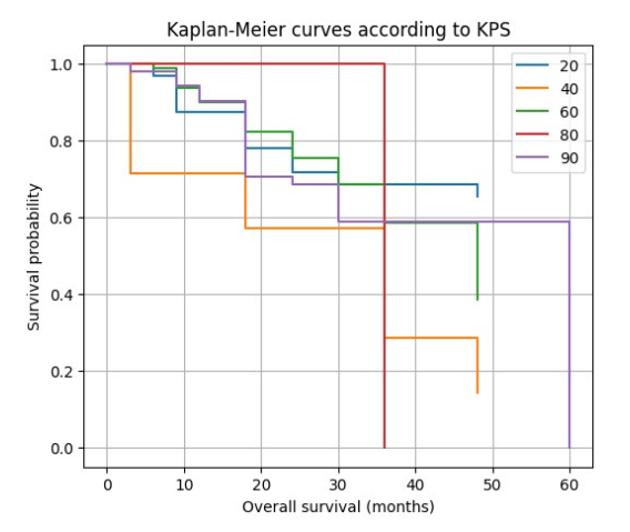
Figure 3: Kaplan–Meier curves according to Karnofsky Performance Status.

Figure 4: Kaplan–Meier curves according to extent of tumor reduction.
Analysis according to radiotherapy technique revealed no statistically significant differences in overall survival among patients treated with 3D-CRT, IMRT, and VMAT [Figure 5], although a tendency toward improved survival was observed in patients treated with modern conformal techniques.

Figure 5: Kaplan–Meier curves according to radiotherapy technique.
These findings suggest that histological grade represents the principal determinant of overall survival, whereas performance status and extent of tumor reduction may also influence long-term outcomes.
Multivariable Cox regression analysis
To identify independent prognostic factors associated with overall survival, a multivariable Cox proportional hazards model was constructed including age, sex, Karnofsky Performance Status, WHO grade, extent of tumor reduction, and radiotherapy technique.
Higher WHO grade was identified as the only independent predictor of worse overall survival. Patients with high-grade tumors had a significantly increased risk of death (HR=1.72, 95% CI 1.06–2.78, p=0.028).
Age (p=0.543), sex (p=0.744), Karnofsky Performance Status (p=0.723), extent of tumor reduction (p=0.122), and radiotherapy technique (p=0.370) were not independently associated with overall survival.
The results of multivariable Cox regression are presented in [Table 5] and [Figure 6].
| Variable | HR | 95% CI | p-value |
|---|---|---|---|
| Age | 0.995 | 0.981–1.010 | 0.543 |
| Sex | 1.08 | 0.69–1.68 | 0.744 |
| KPS | 1.00 | 0.99–1.01 | 0.723 |
| WHO grade | 1.72 | 1.06–2.78 | 0.028 |
| Extent of tumor reduction | 0.99 | 0.99–1.00 | 0.122 |
| Radiotherapy technique | 0.84 | 0.57–1.24 | 0.370 |
Table 5: Multivariable Cox proportional hazards analysis for overall survival.
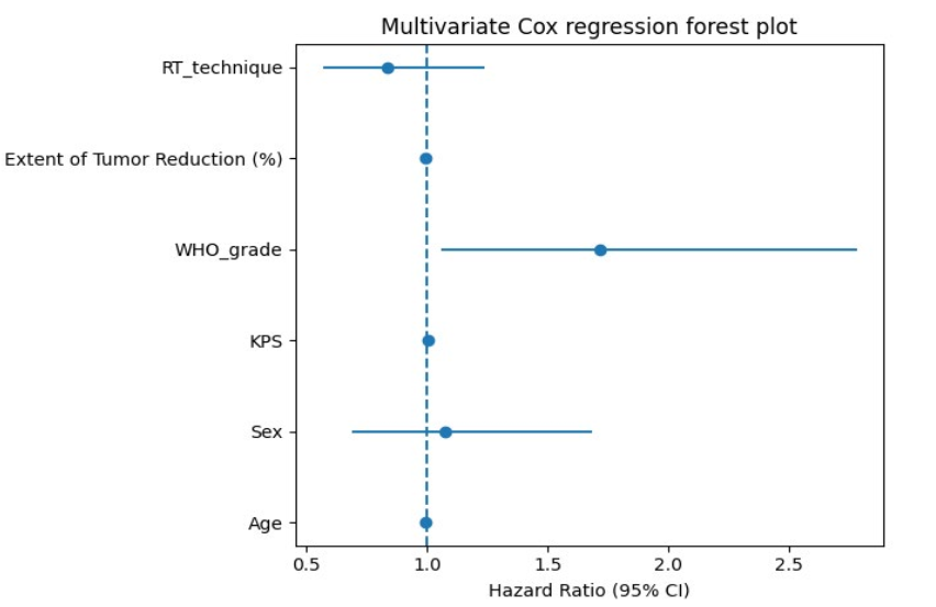
Figure 6: Forest plot of multivariable Cox regression analysis.
DISCUSSION
The present retrospective cohort study characterized neurological manifestations, treatment patterns, postoperative residual disease, and survival outcomes among patients with gliomas treated with surgery followed by postoperative radiotherapy in routine clinical practice in Central Asia. The main findings were that neurological manifestations at presentation were associated with anatomical tumor localization, histological grade was associated with overall survival, and postoperative residual disease was frequently documented. The study also demonstrated substantial heterogeneity in systemic treatment and incomplete availability of molecular and immunohistochemical information.
Neurological manifestations and tumor localization
Neurological symptoms were frequent at presentation, with headache, general weakness, dizziness, seizures, and nausea among the most commonly documented manifestations. The observed association between neurological manifestations and tumor localization is biologically plausible because clinical presentation may vary according to the anatomical structures involved, tumor size, mass effect, and the involvement of eloquent brain regions. The association observed in our cohort was statistically significant and showed a moderate effect size.
These findings are broadly consistent with previous literature describing headache, seizures, cognitive impairment, and focal neurological deficits as common manifestations of gliomas [7]. However, the present study should be interpreted primarily as a description of the clinical presentation of patients treated in routine practice rather than as evidence that tumor localization directly determines the severity of neurological symptoms. The retrospective design did not allow standardized prospective neurological assessment or quantitative measurement of symptom severity.
An important methodological consideration is that neurological manifestations were documented before initiation of postoperative radiotherapy. Therefore, these findings represent baseline disease manifestations and should not be interpreted as treatment-related toxicity.
Histological grade and overall survival
Histological grade was significantly associated with overall survival in the present cohort. Kaplan–Meier analysis demonstrated shorter survival among patients with higher-grade tumors, and histological grade remained associated with mortality in multivariable Cox regression.
This observation is consistent with the established relationship between tumor aggressiveness and survival in glioma populations [11,12,16,22]. However, the finding itself should not be considered a novel biological observation. Rather, its relevance in the present study lies in demonstrating the prognostic pattern within a real-world Central Asian cohort treated over a six-year period.
The interpretation of histological grade requires particular caution in the context of contemporary glioma classification. The 2021 WHO classification integrates histological and molecular characteristics, and molecular alterations such as IDH mutation and 1p/19q codeletion are central to the classification of diffuse gliomas [3]. In our cohort, however, molecular testing was incomplete. IDH status was available only for a minority of patients, while Ki-67 and other immunohistochemical or molecular markers were also unavailable in substantial proportions of the cohort. Consequently, the present analysis should not be interpreted as a comprehensive WHO 2021 molecular classification of all 174 patients.
The observed association between histological grade and survival therefore reflects the classification available in the retrospective institutional records. Molecular heterogeneity may have contributed to the observed survival differences but could not be adequately assessed in the present dataset.
Surgical resection and residual disease
Subtotal resection was the most frequently documented surgical approach, and postoperative MRI demonstrated residual tumor in 156 of 174 patients (89.7%). This high prevalence of residual disease may reflect the anatomical complexity of gliomas, involvement of eloquent structures, tumor location, and the principle of preserving neurological function during surgery.
Patients with greater tumor reduction showed a tendency toward longer survival in the present cohort; however, extent of tumor reduction did not remain independently associated with overall survival in the multivariable model. This finding should be interpreted cautiously because the decision regarding the extent of resection is influenced by tumor location, resectability, functional anatomy, patient condition, and other clinical factors.
Current neuro-oncology recommendations emphasize maximal safe resection when technically feasible [5]. The present findings are compatible with this principle but do not establish a causal relationship between the extent of resection and survival.
The high frequency of residual disease also highlights the importance of postoperative MRI for documenting the extent of residual tumor. Nevertheless, the retrospective nature of the study and the lack of a uniformly documented volumetric assessment limit the precision with which residual tumor burden can be compared between patients.
Radiotherapy and systemic treatment
All patients in the present cohort underwent postoperative radiotherapy, using 3D-CRT, IMRT, or VMAT with prescribed doses ranging from 50 to 60 Gy. The distribution of radiotherapy techniques reflects changes in treatment practice and differences in anatomical and technical requirements over the study period.
An important consideration is that radiotherapy technique was not randomly assigned. In our cohort, radiotherapy technique was associated with tumor localization, indicating potential confounding by anatomical and clinical characteristics. Therefore, differences between treatment techniques should not be interpreted as evidence that one radiotherapy technique is superior to another.
Systemic treatment also varied across the cohort. Temozolomide was the principal systemic agent, while a smaller number of patients received other agents, including irinotecan, lomustine, or bevacizumab. A substantial subgroup did not receive concurrent systemic treatment because of factors documented in the medical records, including less aggressive histology, poor performance status, comorbidities, contraindications, or patient refusal.
This treatment heterogeneity is important when interpreting survival outcomes. Patients receiving or not receiving systemic treatment were unlikely to be clinically equivalent at baseline, creating potential confounding by indication. Accordingly, the present study should be viewed as an analysis of real-world treatment patterns rather than a comparative effectiveness study of radiotherapy or systemic therapy.
Post-treatment neurological findings and adverse events
A substantial proportion of patients had neurological symptoms documented during follow-up, including headache, seizures, memory impairment, and cognitive dysfunction. However, these findings should not automatically be classified as radiation-induced toxicity.
Patients with gliomas may experience persistent or progressive neurological symptoms because of residual disease, disease progression, postoperative changes, seizures, medications, edema, or other neurological factors. In addition, the retrospective medical records did not provide standardized prospective toxicity grading, uniform baseline neuropsychological testing, or systematic evaluation of alternative causes for each neurological finding.
Therefore, the post-treatment findings in this study are more appropriately interpreted as **documented post-treatment clinical events** rather than definitively radiation-attributable complications. This distinction is particularly important because several of the same neurological manifestations were already present before radiotherapy.
Future prospective studies should incorporate standardized toxicity criteria, baseline and longitudinal neurological assessments, cognitive testing, medication documentation, and standardized radiological criteria to distinguish treatment toxicity from progression and other causes.
Disease progression and recurrence
Follow-up MRI demonstrated residual tumor, disease progression, recurrence, and other radiological changes in a proportion of patients. These findings illustrate the clinical complexity of longitudinal imaging assessment after surgery and radiotherapy.
However, postoperative and post-treatment MRI abnormalities may reflect several different processes, including residual tumor, true progression, postoperative change, treatment-related enhancement, pseudoprogression, or radiation necrosis. Because standardized RANO criteria were not consistently documented in the retrospective records, the radiological findings in this study should be interpreted as documented follow-up abnormalities rather than as a definitive standardized classification of progression or recurrence.
This limitation is particularly relevant when interpreting recurrence-related analyses. Prospective studies should use predefined radiological criteria and record the exact dates of progression, recurrence, subsequent treatment, and death to allow robust PFS analysis.
Molecular characterization
The incomplete availability of molecular information represents one of the most important limitations of the present study. IDH testing was available only in a minority of patients, while Ki-67 and other molecular or immunohistochemical markers were also incompletely assessed.
Molecular alterations such as IDH mutation, MGMT promoter methylation, and 1p/19q codeletion have important diagnostic and prognostic implications in contemporary neuro-oncology [3,16,17,23]. Their limited availability prevented meaningful incorporation into the multivariable survival analysis.
Consequently, the present findings should not be generalized to molecularly defined glioma subgroups. Rather, they provide real-world clinical and survival data from a cohort in which molecular characterization was incompletely available. This limitation also emphasizes the need to improve access to standardized molecular diagnostics in future regional studies.
Strengths of the study
Several strengths should be considered. First, the study included 174 consecutive patients treated in routine clinical practice over a defined six-year period. Second, the dataset incorporated demographic, neurological, radiological, surgical, pathological, and treatment-related variables. Third, survival was evaluated using Kaplan–Meier methods and multivariable Cox regression. Finally, the study provides clinical data from a Central Asian population that remains underrepresented in the international neuro-oncology literature.
Limitations
The study has several important limitations. First, its retrospective design introduces the possibility of selection and information bias. Second, the cohort included patients with heterogeneous histological grades and incomplete molecular characterization, limiting the applicability of contemporary molecular classification.
Third, systemic treatment was heterogeneous, and treatment selection was influenced by clinical characteristics. Fourth, radiotherapy technique was associated with tumor localization, raising the possibility of confounding by indication. Fifth, neurological outcomes and post-treatment adverse events were based on retrospective clinical documentation rather than standardized prospective toxicity assessments.
Sixth, postoperative residual disease was assessed from routine MRI documentation rather than from a uniformly performed volumetric analysis. Seventh, the interpretation of progression and recurrence was limited by the absence of consistently documented RANO criteria. Finally, molecular and immunohistochemical variables had substantial missingness and therefore could not be reliably incorporated into the multivariable survival model.
These limitations should be considered when interpreting the associations identified in the present study.
Clinical implications
The findings support the importance of integrating neurological status, anatomical tumor characteristics, surgical findings, pathological grade, and treatment information when evaluating patients with gliomas undergoing postoperative radiotherapy. The high prevalence of residual disease and the frequency of neurological findings during follow-up also underline the importance of structured postoperative imaging and longitudinal neurological assessment.
For future regional studies, standardized molecular classification, systematic documentation of systemic therapy, prospective toxicity assessment, and RANO-based longitudinal imaging would substantially strengthen the evaluation of prognostic factors and treatment outcomes.
CONCLUSIONS
In this retrospective Central Asian cohort of patients with gliomas treated with surgery followed by postoperative radiotherapy, neurological manifestations at presentation were common and were significantly associated with anatomical tumor localization.
Histological grade was significantly associated with overall survival and remained an independent prognostic factor within the multivariable Cox model used in this study. Greater tumor reduction showed a tendency toward improved survival but was not independently associated with overall survival after adjustment for the variables included in the model.
Postoperative residual tumor was frequently documented, occurring in 156 of 174 patients (89.7%). Radiotherapy and systemic treatment patterns were heterogeneous, reflecting differences in clinical and anatomical characteristics.
Neurological symptoms documented during follow-up were frequent; however, given the retrospective design, incomplete baseline assessments, and absence of standardized toxicity grading, these findings should not be interpreted as definitively radiation-induced toxicity.
Finally, incomplete molecular characterization, particularly the limited availability of IDH and other molecular markers, restricts interpretation according to contemporary molecular glioma classification. Prospective multicenter studies incorporating comprehensive molecular profiling, standardized toxicity assessment, RANO-based imaging evaluation, and detailed systemic treatment data are warranted to validate and extend these findings.
Abbreviations
3D-CRT: Three-dimensional conformal radiotherapy
CI: Confidence interval
Gy: Gray
HR: Hazard ratio
IDH: Isocitrate dehydrogenase
IMRT: Intensity-modulated radiotherapy
KPS: Karnofsky Performance Status
MRI: Magnetic resonance imaging
OS: Overall survival
VEGF: Vascular endothelial growth factor
VMAT: Volumetric modulated arc therapy
WHO: World Health Organization
DECLARATIONS
ETHICS APPROVAL AND CONSENT TO PARTICIPATE
The study was approved by the Ethics Committee of Tashkent State Medical University and was conducted in accordance with the Declaration of Helsinki. Due to the retrospective observational design of the study and the use of anonymized clinical and imaging data, the requirement for informed consent was waived by the Ethics Committee.
CONSENT FOR PUBLICATION
Not applicable.
AVAILABILITY OF DATA AND MATERIALS
The datasets generated and/or analyzed during the current study are not publicly available due to institutional and ethical restrictions related to patient confidentiality but are available from the corresponding author on reasonable request.
COMPETING INTERESTS
The authors declare that they have no competing interests.
FUNDING
The authors received no specific funding for this study.
AUTHORS' CONTRIBUTIONS
N.S.K. conceived and designed the study. N.S.K., K.B.A., and A.P.T. collected the clinical and radiological data. D.A.N. performed the histopathological evaluation. Y.M.M. contributed to data interpretation. K.K.K. participated in data collection and manuscript revision. M.R.K. performed the statistical analysis, drafted the manuscript, and served as the corresponding author. All authors critically revised the manuscript and approved the final version.
ACKNOWLEDGEMENTS
Not applicable.
REFERENCES
-
Cramer CK, McKee N, Case LD, Chan MD, Cummings TL, et al. Mild cognitive impairment in long-term brain tumor survivors following brain irradiation. Journal of Neuro-Oncology. 2019;141:235–244. Crossref, Google Scholar, [PubMed]
-
Karimova NS, Nishanova SKh, Yarmatova MB, Musaev DB, Ibragimova GKh. The integration of methods for defining the clinical target volume in radiation therapy for glioblastoma. Journal of Theoretical and Clinical Medicine. 2024;(4):151–154. Google Scholar
-
Louis DN, Perry A, Wesseling P, Brat DJ, Cree IA, et al. The 2021 WHO Classification of Tumors of the Central Nervous System: a summary. Neuro-Oncology. 2021;23:1231–1251. Crossref, Google Scholar, [PubMed]
-
Nishanova YuKh, Karimova NS, Khodjamova GA, Musulmonov ShR. Multiparametric magnetic resonance imaging in the diagnosis of glial brain tumors (review). Journal of Theoretical and Clinical Medicine. 2022;(2):43–50. Google Scholar
-
Niyazi M, Andratschke N, Bendszus M, Chalmers AJ, Erridge SC, et al. ESTRO-EANO guideline on target delineation and radiotherapy details for glioblastoma. Radiotherapy and Oncology. 2023;184:109663. Crossref, Google Scholar, [PubMed]
-
Price M, Ballard C, Benedetti J, Neff C, Cioffi G, et al. CBTRUS Statistical Report: Primary Brain and Other Central Nervous System Tumors Diagnosed in the United States in 2017–2021. Neuro-Oncology. 2024;26:vi1–vi85. Crossref, Google Scholar, [PubMed]
-
Schaff LR, Mellinghoff IK. Glioblastoma and Other Primary Brain Malignancies in Adults: A Review. JAMA. 2023;329:574–587. Crossref, Google Scholar, [PubMed]
-
Stupp R, Mason WP, van den Bent MJ, Weller M, Fisher B, et al. Radiotherapy plus concomitant and adjuvant temozolomide for glioblastoma. N Engl J Med. 2005;352:987–996. Crossref, Google Scholar, [PubMed]
-
van den Bent MJ, Weller M, Wen PY, et al. Clinical perspective on the 2021 WHO CNS tumor classification. Lancet Oncol. 2023;24:e333–e342. Google Scholar
-
van Kessel E, Baumfalk AE, van Zandvoort MJE, Robe PA, Snijders TJ. Tumor-related neurocognitive dysfunction in patients with diffuse glioma: a systematic review of neurocognitive functioning prior to anti-tumor treatment. Journal of Neuro-Oncology. 2017;134:9–18. Crossref, Google Scholar, [PubMed]
-
Weller M, van den Bent M, Preusser M, et al. EANO guidelines on the diagnosis and treatment of diffuse gliomas. Nat Rev Clin Oncol. 2021;18:170–186. Crossref, Google Scholar, [PubMed]
-
Wen PY, Weller M, Lee EQ, Alexander BM, Barnholtz-Sloan JS, et al. Glioblastoma in adults: a Society for Neuro-Oncology (SNO) and European Society of Neuro-Oncology (EANO) consensus review on current management and future directions. Neuro-Oncology. 2020;22:1073–1113. Crossref, Google Scholar, [PubMed]
-
Minniti G, Niyazi M, Andratschke N, et al. Current status and recent advances in reirradiation of glioblastoma. Radiotherapy and Oncology. 2021;157:36–44. Crossref, Google Scholar, [PubMed]
-
Delgado-López PD, Corrales-García EM. Survival in glioblastoma: a review on the impact of treatment modalities. Clinical and Translational Oncology. 2016;18:1062–1071. Crossref, Google Scholar, [PubMed]
-
Ostrom QT, Patil N, Cioffi G, et al. CBTRUS Statistical Report: Primary Brain and Other Central Nervous System Tumors Diagnosed in the United States in 2016–2020. Neuro-Oncology. 2023;25:i1–i99. Crossref, Google Scholar, [PubMed]
-
Molinaro AM, Taylor JW, Wiencke JK, Wrensch MR. Genetic and molecular epidemiology of adult diffuse glioma. Nature Reviews Neurology. 2019;15:405–417. Crossref, Google Scholar, [PubMed]
-
Louis DN, Perry A, Reifenberger G, von Deimling A, Figarella-Branger D, et al. The 2016 World Health Organization Classification of Tumors of the Central Nervous System: a summary. Acta Neuropathologica. 2016;131:803–820. Crossref, Google Scholar, [PubMed]
-
Pegg EC, Zuckerman SL, et al. Seizure management and quality of life in glioma patients. Frontiers in Oncology. 2022;12:849341. Google Scholar
-
Brown TJ, Brennan MC, Li M, Church EW, Brandmeir NJ, et al. Association of the extent of resection with survival in glioblastoma. JAMA Oncology. 2016;2:1460–1469. Crossref, Google Scholar, [PubMed]
-
Stummer W, Pichlmeier U, Meinel T, Wiestler OD, Zanella F, et al. Fluorescence-guided surgery with 5-aminolevulinic acid for resection of malignant glioma. Lancet Oncology. 2006;7:392–401. Crossref, Google Scholar, [PubMed]
-
Ahluwalia MS, Chang SM. Medical therapy for gliomas: current and emerging treatments. Seminars in Neurology. 2018;38:37–46. Google Scholar
-
Weller M, Wick W, Aldape K, Brada M, Berger M, et al. Glioma. Nature Reviews Disease Primers. 2015;1:15017. Crossref, Google Scholar
-
Reifenberger G, Wirsching HG, Knobbe-Thomsen CB, Weller M. Advances in the molecular genetics of gliomas. Nature Reviews Clinical Oncology. 2017;14:434–452. Crossref, Google Scholar, [PubMed]
-
van den Bent MJ, Brandes AA, Taphoorn MJB, Kros JM, Kouwenhoven MC, et al. Adjuvant procarbazine, lomustine, and vincristine chemotherapy in newly diagnosed anaplastic oligodendroglioma. Lancet. 2013;382:1044–1052. Crossref, Google Scholar, [PubMed]
-
Chinot OL, Wick W, Mason W, Henriksson R, Saran F, et al. Bevacizumab plus radiotherapy-temozolomide for newly diagnosed glioblastoma. New England Journal of Medicine. 2014;370:709–722. Crossref, Google Scholar, [PubMed]