Research Article - Onkologia i Radioterapia ( 2026) Volume 20, Issue 8
End-to-end dosimetric evaluation of ITV margins in lung SBRT using a QUASAR phantom
Adeb A.S.A. Almaamari1,2*, K. Nouni1,2, A. Ait Errouhi1, M.A. Youssoufi1, M. Driouch3, A. Lachgar1,2, H. Elkacemi1,2, T. Kebdani1,2 and K. Hassouni1,22Department of Medicine, Mohammed V University, Rabat, Morocco
3LPMS, Faculty of Sciences, Ibn Tofail University, Kenitra, Morocco
Adeb A.S.A. Almaamari, Department of Radiotherapy, National Institute of Oncology, UHC Ibn Sina, Rabat, Morocco, Email: adeebalmamari66@gmail.com
Received: 08-Jun-2026, Manuscript No. OAR-26-194802; , Pre QC No. OAR-26-194802 (PQ); Editor assigned: 10-Jun-2026, Pre QC No. OAR-26-194802 (PQ); Reviewed: 24-Jun-2026, QC No. OAR-26-194802; Revised: 10-Aug-2026, Manuscript No. OAR-26-194802 (R); Published: 17-Aug-2026
Abstract
Background: Stereotactic Body Radiotherapy (SBRT) is an advanced technique for treating lung cancer with high precision, but respiratory-induced tumor motion remains a major challenge. To assess the quality and safety of SBRT treatments through an end-to-end test, ensuring treatment reproducibility, accuracy, and patient safety.
Materials and methods: Respiratory tumor motion was simulated using QUASAR and Octavius 4D phantoms, with 4DCT-based SBRT planning using Monaco 5.1. Dosimetric verification was performed using the Anzai system, Semiflex 3D ionization chamber, and Octavius 4D 1600 SRS array.
Results: Based on the results obtained, all measured relative dose deviations remained within the acceptable ± 5% limit recommended by ICRU TG-142, confirming the dosimetric accuracy and reliability of the treatment plans. The 3-arc VMAT plans demonstrated the best overall dosimetric conformity, particularly under irregular and rapid respiratory cycles. Furthermore, the Gamma Index analysis showed excellent agreement (>90%) between measured and calculated doses across all respiratory conditions, supporting the robustness and clinical reliability of the studied plans.
Conclusion: This end-to-end SBRT study demonstrated good agreement between planned and delivered doses across regular, irregular, and rapid respiratory patterns, with both VMAT and DCA achieving acceptable dosimetric accuracy and conformity. Increasing the number of arcs reduced dose deviations and mitigated the respiratory Interplay effect, despite target-motion uncertainties caused by baseline shifts and variations in respiratory amplitude and period.
Keywords
Lung SBRT; End-to-end dosimetry; ITV margin; QUASAR phantom; Respiratory motion; Dosimetric verification; Interplay effect
Introduction
Stereotactic Body Radiotherapy (SBRT) enables highly precise treatment of lung cancer; however, respiratory-induced tumor motion remains a critical challenge that can affect treatment accuracy and dosimetric reliability, an end-to-end dosimetric test of the Internal Target Volume (ITV) approach applied to Stereotactic Body Radiotherapy (SBRT) for lung cancer was conducted [1,2].
This test to reproduce the full therapeutic process, from treatment simulation to final administration, in order to evaluate the robustness and reliability of the ITV strategy under simulated clinical conditions. To do this, we used a dynamic QUASAR phantom, capable of reproducing different respiratory patterns, including regular breaths to slightly irregular breaths, as well as highly disturbed breaths in terms of period and amplitude. This diversity of respiratory scenarios allowed us to test the system's performance under conditions close to clinical reality, where tumor movement induced by respiration constitutes a major factor of uncertainty, a complete dosimetric verification was carried out to compare and analyse four types of treatment planning. Two plans were designed without collimator rotation: one plan in Volumetric Modulated Arc Therapy (VMAT) and one plan in Dynamic Conformal Arc (DCA). In parallel, two other plans were developed by integrating three collimator rotations, one in VMAT and the other in DCA, in order to evaluate the impact of this strategy on dosimetric accuracy and conformity. The analysis focused on the three studied respiratory cycles, allowing for the measurement of the discrepancies between the planned doses and the doses actually delivered under free-breathing conditions [3].
This experimental protocol not only validated the relevance of the ITV approach in the context of pulmonary SBRT but also demonstrated the advantages and limitations of different planning techniques. The use of the QUASAR phantom provided a reliable platform to simulate tumor movement and analyse the effect of the respiratory cycle on dose distribution. The results obtained demonstrate the importance of appropriate planning, particularly through the increase in the number of arcs and the optimization of collimation parameters, in order to reduce uncertainties related to respiratory movement and improve the robustness of the treatment [4].
This end-to-end test thus constitutes an essential step in the quality assurance of SBRT treatments, ensuring better reproducibility and increased safety for patients.
Materials and Methods
Phantoms QUASAR pRESP, Octavuis SRS 1600 4D
The QUASAR phantom is a programmable respiratory motion simulator designed to ensure the quality of motion-guided treatments in radiotherapy. It simulates complex respiratory movements, including superior/inferior translation and optional lateral hysteresis, and is specifically designed for IGRT (Image- Guided Radiation Therapy) and SGRT (Surface Guided Radiation Therapy) techniques. The QUASAR allows you to choose between simple operation with local manual control at the motor level and advanced programmable control driven by software. It allows reading the movement waveforms, which enables testing the limits of motion management systems. It is in the form of an oval body (30(L) × 20(W) × 12(H) cm³) with a density of 1.20 g/ cm³, equivalent to that of a thorax, and an opening allowing the insertion of a hollow cedar cylindrical insert that contains a sphere with a diameter of 30 mm, considered a tumor. Linked to a training unit that allows creating, modifying, and recording movement waveforms and sending these commands to the phantom.
To evaluate the impact of various density overrides on lung SBRT plans, a phantom study was conducted using the Elekta synergy linear accelerator and the Monaco 5.1 planning system. A programmable QUASAR respiratory motion phantom (Modus Medical Devices Inc., London, ON, Canada) with a cork insert containing a hollow sphere considered the target tumor was used to simulate the craniocaudal motion of a pulmonary tumor in an SBRT patient.
The scanner dedicated to the radiotherapy department of the National Institute of Oncology INO, a Siemens SOMATOM® Definition CT scanner (Siemens Healthcare Limited). This scanner was used to acquire 4DCT scans with the QUASAR phantom set to a maximum respiratory period of 3.8 seconds and a maximum motion amplitude of about 0.6 inches. The respiratory waveform was recorded simultaneously with the Anzai respiratory control system. For each of the different respiratory cycles, the reconstruction process generated the Average Intensity Projection (AIP) and a dataset containing each of the 8 respiratory phases from which the Maximum Intensity Projection (MIP) was generated. The three required image datasets (AIP, MIP, and a 50% central phase dataset) were exported to the TPS. The contouring of the two target volumes, the ITV and the PTV, was completed using the Monaco 5.1 planning system (Elekta, Inc) and utilized the MIP of each of the 4D CT scans for the different cycles. The expansion of the PTV was 1 cm in the superior/inferior direction and 0.5 cm in the axial plane. The contours were applied to each of the AIP and central phase datasets at 50% for each cycle (Figures 1 and 2).
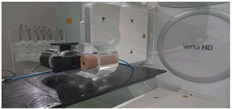
Fig. 1. The QUASAR phantom.
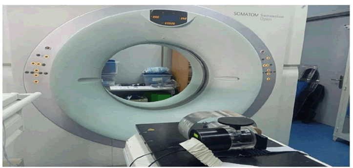
Fig. 2. CT simulator SIEMENS SOMATOM sensation open.
Anzai respiratory control system: The Anzai respiratory control system is a device used in radiotherapy to manage, monitor, and record patients' respiratory movements. It consists of a fixation belt that is used to position a pressure transducer in a patient's chest. Due to the expansion and contraction of the belt during breathing, the pressure from the transducer delivers a digital signal. This signal will be amplified and then evaluated by the Anzai software, thus recording the respiratory cycle. The components of the system: Pressure transducer attached by a fastening belt, fastening belts of different sizes, electrical amplifier (Figure 3).
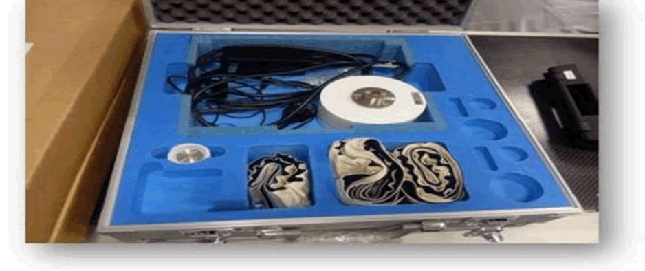
Fig. 3. The ANZAI respiratory cycle recording system.
Semiflex 3D ionization chamber PTW-31021: The Semiflex 3D PTW-31021 ionization chamber is an ionization detector with a sensitive volume of 0.07 cm³, designed for absolute and relative dosimetry measurements in water, air, or solid phantoms. It is equipped with a complete 3D geometry, which means it offers a uniform response in all directions, a minimal sensitive volume that reduces the volume averaging effect, which is particularly useful for dose measurements in small fields [5,6], a low polarity effect, which means the chamber is more accurate and reliable in its measurements, and a short stabilization time.
The Semiflex 3D PTW-31021 is designed to meet the requirements of dosimetry standards, particularly those of AAPM TG-51 [7,8], and is considered an ideal tool for clinical and research applications in dosimetry (Figure 4).
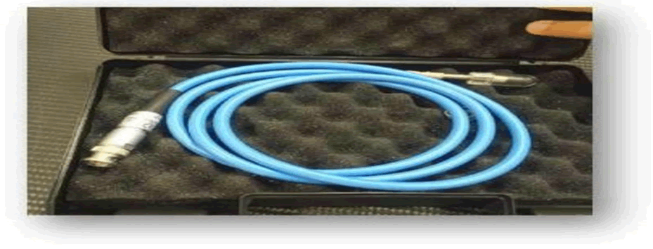
Fig. 4. The semi flex 3D PTW-31021 ionization chamber.
Octavius 1600 SRS 4D: It is a versatile anthropomorphic phantom used in radiotherapy to measure the absorbed dose and evaluate the accuracy of treatment planning, thus allowing for the validation of dosimetry in realistic conditions.
The device includes the Octavius 4D phantom, with a 2D Octavius 1000 SRS matrix of liquid ionization chambers (PTW-Freiburg, Germany). Indeed, during irradiation, the phantom rotates with the linac arm thanks to an inclinometer fixed on it. At each angle, the 1000SRS matrix is perpendicular to the beam. The PTW acquisition software (Mephysto) reconstructs a 3D dose matrix in a homogeneous water-equivalent cylinder using PDDs measured under reference conditions (Figure 5) [9].
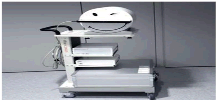
Fig. 5. The Octavius 4D phantom, with a 2D 1600 SRS matrix of liquid ionization chambers.
Versa HD accelerator: The Versa HD linear accelerator is a device marketed by Elekta, dedicated to external radiotherapy treatments and capable of delivering electron or photon beams with energies ranging from 4 to 25 MeV. It allows for conventional radiotherapy treatments, intensity-modulated (IMRT, VMAT), as well as highdose- rate stereotactic treatments (FFF), with stereotactic technology that locates and treats primary or metastatic tumors. The system is equipped with a robotic positioning table, an Agility Multi-Leaf Collimator (MLC) to precisely control the shape and size of the radiation field, and offers options for respiratory compensation and on-board imaging systems to improve patient positioning accuracy
TPS Monaco: The TPS Monaco Version 5.11.03 is a radiation therapy planning system developed by Elekta, used to generate high-performance and precision treatment plans for all 3D radiation therapy techniques, IMRT, and VMAT. It uses Monte Carlo and collapsed cones algorithms to calculate plans while ensuring excellent dose distribution, maximizing the sparing of healthy tissues, and improving treatment efficiency. Monaco is also compatible with the most innovative radiotherapy techniques. The system also allows the import and comparison of treatment plans generated by other TPS to perform quality control of treatment plans [10].
Patient selection and contouring and planning process
Choice of respiratory cycles: For this study, three distinct respiratory cycles were selected from the QUASAR™ respiratory motion application, using an advanced programmable control controlled by software. This application allows for the generation and configuration of respiratory cycles by adjusting either the amplitude of the cycle, its period, or the number of cycles per minute (BPM). The first cycle thus chosen corresponds to a regular, calm, and constant breathing, characterized by an amplitude of 1.5 cm and a period of 3.8 seconds. The second cycle simulates a more erratic and less regular breathing, with an amplitude of less than 1.5 cm and a variable period. Finally, the third cycle represents a stressed and faster breathing, with a variable amplitude and period. This variation in respiratory cycles aims to take into account the different types of breathing that patients may adopt, whether during simulation periods or during treatment (Figure 6) [11].
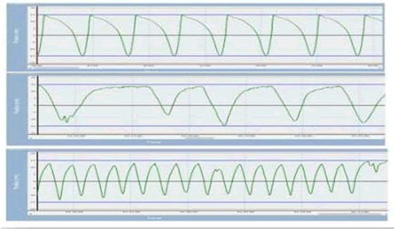
Fig. 6. Regular, irregular and fast respiratory cycles.
Recording of respiratory cycles by 4DCT: The respiratory cycles thus defined will be captured by a 4DCT tomographic acquisition, performed with the SIEMENS SOMATOM Sensation scanner and ensured by the Anzai Medical brand recording system. This ANZAI system records the amplitude and shape of the respiratory cycle, then divides it into eight phases, 4 phases during the inspiration period (0%, 25%, 75%, 100%) and 4 phases during expiration (0%, 25%, 50%, 75%).
The acquired images are then classified according to the corresponding respiratory phase. Each phase is identified as GTV, indicating the position of the tumor during respiration, and marked in its reference scan (CT0%, CT25%, CT50%, CT100%) during inspiration and the others during expiration (CT 0%; CT 25%; CT 50%; CT 75%) (Figure 7) [12].
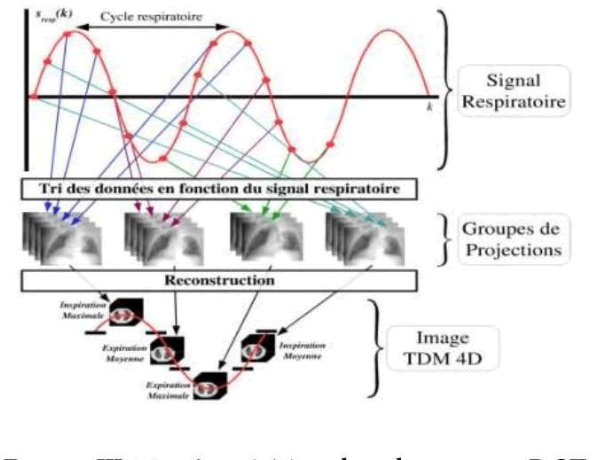
Fig. 7. Phase acquisition by 4DCT.
The acquisition of slices for all cycles was performed in helical mode, using identical parameters, namely a slice thickness of 1.5 mm, a pitch of 1, and a FOV (Field of View) of 350 mm. The CTs of the eight phases will then be sent to the processing system for delineation and treatment planning [13].
Delimitation of the ITV volume of each respiratory cycle: In our study, we opted for the method based on MIP reconstruction, which was automatically generated using the Monaco planning system available in the department. The MIP image represents, for each voxel, the maximum intensity value observed among all phases of the respiratory cycle. This technique highlights the extreme positions of the tumor during respiration, which is essential for capturing the full range of its movement. Once the MIP reconstruction is completed, the ITV volume is delineated directly on this image. Subsequently, this contouring is merged with another reconstruction, called AVE (Average Intensity Projection). The averaged AVE image, also created on the TPS Monaco, corresponds to an average of the intensities over all respiratory phases. It provides a more stable and homogeneous representation, used as a basis for treatment planning. The combined use of MIP and AVE reconstructions is crucial for optimal consideration of tumor movements related to breathing: The MIP image ensures that the ITV volume encompasses all possible positions of the tumor, while the AVE image allows for precise and realistic dose planning (Figure 8) [14-16].
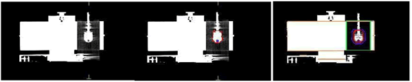
Fig. 8. Reconstruction in MIP, contoured MIP and AVE contoured.
Contour of structures: Two main structures have been created for treatment planning: The PTV volume defined with a 5 mm margin around the ITV to account for localization uncertainties, and the PTV – ITV volume, representing the difference between the PTV and ITV. An optimal window of 400 units Hounsfield was used for contouring the structures of interest, which include the external contour, the insert reproducing respiratory movements, the volume representing the tumor, the ITV encompassing all positions of the tumor, the ionization chamber, the PTV, and the PTV – ITV volume (Figure 9).
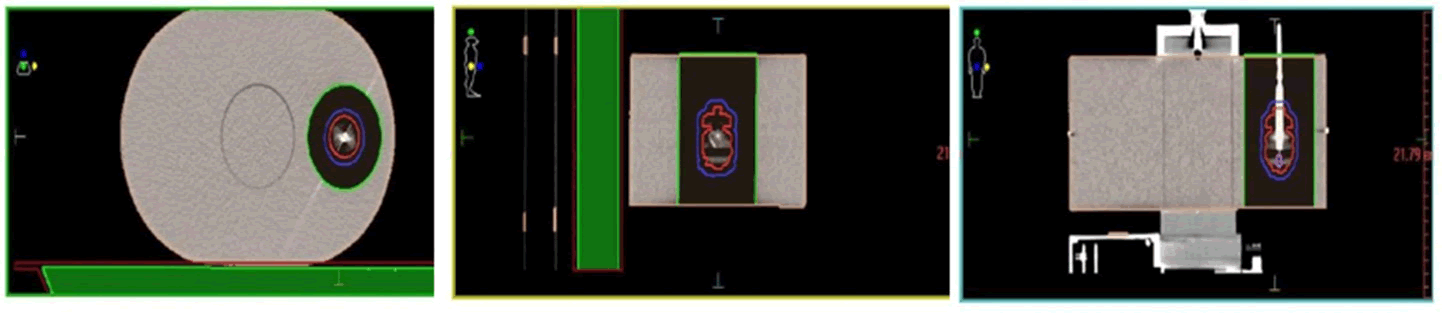
Fig. 9. Axial, sagittal, and coronal sections
Planning in Monaco: The treatment planning was carried out using the TPS Monaco version 5.11 planning system, with calculations performed using the Monte Carlo algorithm, the latter used a calculation grid of 1.5 mm to ensure optimal precision in dose distribution. The prescribed dose for each treatment is 8 Gy, administered in a single fraction. For each respiratory cycle, four coplanar SBRT (Stereotactic Body Radiation Therapy) treatment plans were designed, using a 6MV FFF (Flattening Filter Free) beam [17].
These plans are divided into two distinct planning techniques:
• VMAT (Volumetric Modulated Arc Therapy) plan without collimator rotation.
• VMAT plan with three collimator rotations at angles of 0°, 20°, and 340°.
• DCA (Dynamic Conformal Arc) plan without collimator rotation.
• DCA plan with three collimator rotations at angles of 0°, 20°, and 340°.
Results
Dosimetric evaluation
The results obtained for the relative dose deviations were analysed according to the different types of respiratory cycles studied. For the regular cycle, the observed deviations are -2.315% for the plan with 1 VMAT arc, 0.0574% for 3 VMAT arcs, -2.488% for 1 DCA arc, and -2.2577% for 3 DCA arcs. Regarding the irregular cycle, the deviations are -3.35% for 1 VMAT arc, - 1.548% for 3 VMAT arcs, -2.835% for 1 DCA arc, and -3.360% for 3 DCA arcs. Finally, for the fast cycle, the measured deviations are -2.9% for 1 VMAT arc, -1.52% for 3 VMAT arcs, - 4.273% for 1 DCA arc, and -1.33% for 3 DCA arcs.
According to the recommendations of the ICRU TG 142 report, the relative dose deviation should not exceed 5%. These results remain within the acceptable limits defined by the ICRU recommendations.
All measured deviations remain within the acceptable limit of ±5% defined by the ICRU, thus confirming the quality and accuracy of the studied plans. The results show better conformity of the 3-arc VMAT plans compared to other configurations, particularly in irregular and rapid cycles (Tables 1 and 2).
|
|
Calculated dose (Gy) |
Measured dose (Gy) |
Relative error (%) |
|
Arc Vmat |
8,63 |
8,43 |
-2,315 |
|
Arcs Vmat |
9,165 |
9,17 |
0,05 |
|
Arc DCA |
10,047 |
9,797 |
-2,488 |
|
Arcs DCA |
10,035 |
9,808 |
-2,25 |
Tab. 1. Calculated and measured doses for the regular cycle.
|
|
Calculated dose (Gy |
Measured dose (Gy) |
Relative error (%) |
|
Arc Vmat |
9,555 |
9,274 |
-2,94 |
|
Arcs Vmat |
9,283 |
9,176 |
-1,15 |
|
Arc DCA |
9,97 |
9,544 |
-4,27 |
|
Arcs DCA |
8,741 |
8,624 |
-1,337 |
Tab. 2. Calculated and measured doses for the fast cycle.
Gamma index analysais
|
|
Gamma index analysais |
Collimator angle |
Prescribed dose |
|
|
2%/2 mm |
3%/2 mm |
|||
|
Arc Vmat |
97,20% |
98,70% |
0° |
8Gy/fraction |
|
Arcs Vmat |
95,20% |
98,20% |
0°, 20°, 340° |
8Gy/fraction |
|
Arc DCA |
98,30% |
99,60% |
0° |
8Gy/fraction |
|
Arcs DCA |
95,50% |
98,10% |
0°, 20°, 340° |
8Gy/fraction |
Tab. 3. Gamma analysis table 2%/2 mm and 3%/2 mm for the regular cycle.
|
|
Gamma index analysis |
Collimator angle |
Prescribed dose |
|
|
2%/2 mm |
3%/2 mm |
|||
|
Arc Vmat |
96,20% |
98,30% |
0° |
8Gy/fraction |
|
Arcs Vmat |
93,20% |
97,60% |
0°, 20°, 340° |
8Gy/fraction |
|
Arc DCA |
94,70% |
97,40% |
0° |
8Gy/fraction |
|
Arcs DCA |
94,20% |
97,50% |
0°, 20°, 340° |
8Gy/fraction |
Tab. 4. Gamma analysis table 2%/2 mm and 3%/2 mm for the irregular cycle.
|
|
Gamma index analysis |
Collimator angle |
Prescribed dose |
|
|
2%/2 mm |
3%/2 mm |
|||
|
Arc Vmat |
90,50% |
94,30% |
0° |
8Gy/fraction |
|
Arcs Vmat |
94,20% |
97,40% |
0°, 20°, 340° |
8Gy/fraction |
|
Arc DCA |
93,00% |
96,20% |
0° |
8Gy/fraction |
|
Arcs DCA |
92,20% |
95,50% |
0°, 20°, 340° |
8Gy/fraction |
Tab. 5. Gamma analysis table 2%/2 mm and 3%/2 mm for the fast cycle.
Discussion
The radiotherapy of bronchial tumors must take into account respiratory movement. Indeed, while the target (tumor) is being irradiated, it can move by several centimeters, which can potentially lead to under dosing of the target and overdosing of the organs at risk. For conventional irradiations, this movement is generally taken into account by using safety margins [18]. But, when the treatment is intensity-modulated V mat, there is an additional risk of an unfortunate combination of collimator leaf movement and respiratory motion: Interplay effect [19,20].
To minimize and mitigate this effect in VMAT stereotactic treatments without flattening filters, Ong et al., showed that the dose discrepancies within the lung due to the interplay effect were significantly reduced when the dose rate was decreased and the number of arcs was increased (from 1 arc to 2 arcs) [21]. Similarly, it is unlikely that this effect is clinically significant for stereotactic lung treatments when using at least two VMAT arcs for treatments involving more than one fraction [22]. Our study showed that increasing the number of arcs in treatment planning significantly reduces the discrepancies between the planned dose and the actual delivered dose, for regular, irregular, and rapid respiratory cycles.
This improvement is even more pronounced in the case of the irregular respiratory cycle, where increasing the number of arcs mitigates the dose variations induced by respiratory movements. For example, for the regular respiratory cycle, the dose deviation changes from -2.315% with a single arc to 0.0574% with three arcs, highlighting a notable improvement in precision.
In the case of the fast cycle, this gap decreases from -2.9% to -1.152% when moving from one to three arcs. For the irregular cycle, the gap decreases from -3.35% to -1.548%, demonstrating the increased effectiveness of this strategy in reducing significant discrepancies. The results obtained in our study are consistent with those of [23], where the authors evaluated the Interplay effect as well as the influence of the number of arcs on dosimetric accuracy in the context of VMAT treatment for the lung. Using a Halcyon accelerator with a 6 MV FFF unfiltered beam and a programmable thoracic motion phantom, they found that the maximum and average dose discrepancies significantly decrease when the number of arcs increases from one to two, with a reduction in the discrepancy from 7.2% to 1.8%. The consistency between the two studies supports the idea that increasing the number of arcs helps reduce the adverse effects related to respiratory movements and collimator movement, while improving the precision of dose distribution in stereotactic treatments for lung tumors. In conclusion, our results, as well as those of the article, support that increasing the number of arcs in planning is an effective strategy to mitigate the impact of respiratory movements, particularly in the case of rapid respiratory cycles, where dose discrepancies are often more pronounced. This optimization is essential for treatments under stereotactic conditions, where high precision is required to deliver the dose while minimizing exposure to healthy tissues.
Currently, Dynamic Conformal Arcs (DCA) and Volumetric Modulated Arc Therapy (VMAT) are two techniques commonly used for the planning of stereotactic lung radiotherapy treatments. DCA offers certain advantages in terms of Multileaf Collimator (MLC) movement and delivery efficiency. However, VMAT is often preferred when the preservation of organs at risk becomes a priority. In our study, we compared these two techniques and found a significant deviation in the dose delivered with DCA across all respiratory cycles. For the regular cycle, the deviation was -2.48%, for the irregular cycle it was -3.36%, and for the fast cycle it was - 4.27%.
In comparison with the article "Comparison of the dosimetric planning efficiency of dynamic conformal arc and volumetric modulated arc therapy techniques for stereotactic body radiotherapy of lung cancer using internal target volume" [3], which used the same free-breathing strategy and ITV creation, the authors concluded that the DCA technique has been clinically validated as a safe, effective, and acceptable treatment compared to the standard VMAT technique in SBRT for non-small cell lung cancer. By reducing the uncertainty of small field dosimetry and the interplay effect, while providing better transition rates, DCA could be used as the first treatment option in clinics without a respiratory cycle management system. DCA achieved excellent plan quality, better preserving healthy lungs and significantly reducing delivery time and dose (MU), thus accelerating treatment delivery. Our results seem to contradict those of this article, as in our study, we observed that VMAT was more effective than DCA. This contradiction can be explained by the fact that we used a single DCA arc, whereas in the mentioned article, two DCA arcs were used, which can further reduce the dose discrepancies caused by the interplay effect. This difference in the number of arcs may explain the variation in the results observed between the two studies.
Treatment planning for pulmonary locations presents specific challenges for dose calculation algorithms in Treatment Planning Systems (TPS). Indeed, the low densities of the tissues that compose and/or surround the target are treated differently depending on the algorithm used. According to the latest recommendations [24], it is advised to avoid type A algorithms, which do not properly account for tissue heterogeneities, and to favor type B or C algorithms, such as those based on Monte Carlo. The latter precisely model the lateral transport of electrons in low-density areas, which allows for better consideration of dose variations in these regions. In our study, we applied a hybrid method to the rapid cycle in order to enhance the density with the aim of improving planning accuracy. This hybrid method involves assigning the average density of the GTV (Gross Tumor Volume) to the ITV (Internal Target Volume) and assigning an intermediate value between the average lung density and that of the ITV to the PTV margin (Planning Target Volume). This approach can be used regardless of the type of treatment technique, whether it be fixed beams, Dynamic Conformal Arc (DCA), static IMRT, or Volumetric Modulated Arc Therapy (VMAT). Our results show that the dose deviations for all techniques (DCA, VMAT) are not significantly different when applying this hybrid method, compared to standard plans without density enhancement [25,26].
These results are significant because the Monte Carlo algorithm used in our TPS automatically adjusts density heterogeneities, making the application of an additional hybrid method during planning unnecessary. The Monte Carlo algorithm effectively corrects these density variations, thereby improving the accuracy of the delivered doses, particularly in structures like the lungs, where density differences are pronounced [27,28].
Conclusion
It is very important to this study; an end-to-end test in SBRT treatment for lung cancer was conducted. The test showed a good concordance between the planned doses and the doses actually delivered, for regular and slightly irregular respiratory patterns (variation in the period of respiration) and rapid (variation in the amplitude and period of respiration). Dose measurements were performed using the anthropomorphic phantom Octavius 4D and the Semi flex 3D ionization chamber. For the respiratory patterns, the measured doses were underestimated due to inappropriate target movement within the predetermined trigger window (unstable target), affected by baseline shifts and variations in respiratory amplitude and period. The results showed that the two treatment techniques studied, VMAT and DCA, present acceptable dose deviations as well as a conformal dose distribution for all the respiratory cycles analysed within the framework of free breathing with the ITV strategy. Furthermore, it was observed that increasing the number of arcs in planning significantly helps reduce deviations, thereby minimizing the interplay effect, which is responsible for overdoses or underdoses.
References
- Ong CL, Dahele M, Slotman BJ, Verbakel WFAR. Dosimetric impact of the interplay effect during stereotactic lung radiation therapy delivery using flattening filter-free beams and volumetric modulated arc therapy. Int J Radiat Oncol Biol Phys. 2013; 86:743-748.
- Edvardsson A, Nordström F, Ceberg C, Ceberg S. Motion induced interplay effects for VMAT radiotherapy. Phys Med Biol. 2018; 63:085012.
[Crossref] [Google Scholar] [PubMed]
- Bertholet J, Knäusl B, Fogliata A. Dynamic conformal arcs for lung stereotactic body radiation therapy: A comparison with volumetric-modulated arc therapy. Med Phys. 2016; 43:1784-1794.
[Crossref] [Google Scholar] [PubMed]
- Almond PR, Biggs PJ, Coursey BM. AAPM's TG-51 protocol for clinical reference dosimetry of high-energy photon and electron beams. Med Phys. 1999; 26:1847-1870.
[Crossref] [Google Scholar] [PubMed]
- McEwen MR, DeWerd LA, Ibbott GS. Addendum to the AAPM's TG-51 protocol for clinical reference dosimetry of high-energy photon beams. Med Phys. 2014; 41:041501.
- Andreo P, Burns DT, Hohlfeld K. Absorbed dose determination in external beam radiotherapy: An international code of practice for dosimetry based on standards of absorbed dose to water. IAEA Technical Reports Series No. 398. Vienna: IAEA; 2000.
- Klein EE, Hanley J, Bayouth J. Task Group 142 report: Quality assurance of medical accelerators. Med Phys. 2009; 36:4197-4212.
[Crossref] [Google Scholar] [PubMed]
- Li XA, Liu C, Ajlouni M. Interplay between breathing and MLC motion in VMAT for lung SBRT: A Monte Carlo simulation study. Med Phys. 2013; 40:111714.
- Underberg RWM, Lagerwaard FJ, Slotman BJ. Benefit of respiration-gated stereotactic radiotherapy for stage I lung cancer: An analysis of 4DCT datasets. Int J Radiat Oncol Biol Phys. 2005; 62:554-560.
[Crossref] [Google Scholar] [PubMed]
- Wolthaus JWH, Schneider C, Sonke JJ. Mid-ventilation CT scan construction from four-dimensional respiration-correlated CT scans for radiotherapy planning of lung cancer patients. Int J Radiat Oncol Biol Phys. 2006; 65:1560-1571.
[Crossref] [Google Scholar] [PubMed]
- Rietzel E, Chen GTY, Choi NC, Willet CG. Four-dimensional image-based treatment planning: Target volume segmentation and dose calculation in the presence of respiratory motion. Int J Radiat Oncol Biol Phys. 2005; 61:1535-1550.
- Keall PJ, Mageras GS, Balter JM. The management of respiratory motion in radiation oncology report of AAPM Task Group 76. Med Phys. 2006; 33:3874-3900.
[Crossref] [Google Scholar] [PubMed]
- Low DA, Moran JM, Dempsey JF, Dong L, Oldham M. Dosimetry tools and techniques for IMRT. Med Phys. 2011; 38:1313-1338.
[Crossref] [Google Scholar] [PubMed]
- Low DA, Harms WB, Mutic S, Purdy JA. A technique for the quantitative evaluation of dose distributions. Med Phys. 1998; 25:656-661.
[Crossref] [Google Scholar] [PubMed]
- Ono T, Ishida T, Yasuda K. Evaluation of the interplay effect in lung stereotactic body radiation therapy using a dynamic thoracic phantom. J Appl Clin Med Phys. 2020; 21:102-111.
- Guckenberger M, Richter A, Krieger T. Is a single respiratory correlated 4D-CT study sufficient for evaluation of breathing motion? Int J Radiat Oncol Biol Phys. 2008; 72:683-690.
[Crossref] [Google Scholar] [PubMed]
- Boda-Heggemann J, Knopf AC, Simeonova A. Deep inspiration breath hold-based radiation therapy: A clinical review. Int J Radiat Oncol Biol Phys. 2016; 94:478-492.
[Crossref] [Google Scholar] [PubMed]
- Underberg RWM, Lagerwaard FJ, Cuijpers JP, Slotman BJ, van Sornsen de Koste JR, et al. Four-dimensional CT scans for treatment planning in stereotactic radiotherapy for stage I lung cancer. Int J Radiat Oncol Biol Phys. 2004; 60:1283-1290.
[Crossref] [Google Scholar] [PubMed]
- Timmerman R, McGarry R, Yiannoutsos C. Excessive toxicity when treating central tumors in a phase II study of stereotactic body radiation therapy for medically inoperable early-stage lung cancer. J Clin Oncol. 2006; 24:4833-4839.
[Crossref] [Google Scholar] [PubMed]
- Timmerman RD, Park C, Kavanagh BD. The North American experience with stereotactic body radiation therapy in non-small cell lung cancer. J Thorac Oncol. 2007; 2:S101-S112.
[Crossref] [Google Scholar] [PubMed]
- Chang JY, Senan S, Paul MA. Stereotactic ablative radiotherapy versus lobectomy for operable stage I non-small-cell lung cancer: A pooled analysis of two randomised trials. Lancet Oncol. 2015; 16:630-637.
- ICRU Report 91 Prescribing, Recording, and reporting of stereotactic treatments with small photon beams. J ICRU. 2016; 16:1-160.
- Das IJ, Cheng CW, Watts RJ. Accelerator beam data commissioning equipment and procedures: Report of the TG-106 of the Therapy Physics Committee of the AAPM. Med Phys. 2008; 35:4186-4215.
[Crossref] [Google Scholar] [PubMed]
- Das IJ, Ding GX, Ahnesjö A. Small fields: Non-equilibrium radiation dosimetry. Med Phys. 2008; 35:206-215.
[Crossref] [Google Scholar] [PubMed]
- Francescon P, Cora S, Satariano N. Calculation of k(Qclin, Qmsr) for several small detectors and for two linear accelerators using Monte Carlo simulations. Med Phys. 2011; 38:6513-6527.
[Crossref] [Google Scholar] [PubMed]
- Benedict SH, Yenice KM, Followill D. Stereotactic body radiation therapy: The report of AAPM Task Group 101. Med Phys. 2010; 37:4078-4101.
[Crossref] [Google Scholar] [PubMed]
- Bortfeld T, Jiang SB, Rietzel E. Effects of motion on the total dose distribution. Semin Radiat Oncol. 2004; 14:41-51.
[Crossref] [Google Scholar] [PubMed]
- Jiang SB, Pope C, Al Jarrah KMsssss. An experimental investigation on intra-fractional organ motion effects in lung IMRT treatments. Phys Med Biol. 2003; 48:1773-1784.
[Crossref] [Google Scholar] [PubMed]