Review Article - Onkologia i Radioterapia ( 2020) Volume 14, Issue 3
The promising applications of ultrasound in emergency medicine and critical care related to in cancer: a review
Samira Vaziri1, Reza Mosaddeghi1, Gholamreza Masoumi2,3, Mehdi Rezai1 and Fatemeh Mohammadi1*2Trauma and Injury Research Center, Tehran, Iran
3Health in Emergency and Disaster Research Center, University of Social Welfare and Rehabilitation Sciences, Tehran, Iran
Fatemeh Mohammadi, Emergency Medicine Management Research Center, Tehran, Iran, Tel: +98-2155228580, Email: dr.fmohammadi304@yahoo.com
Received: 07-Jan-2020 Accepted: 22-Jan-2020 Published: 30-Jan-2020
Abstract
The referral of critically ill cancer patients to an Intensive Care Unit (ICU) is a matter of controversial debate. During the past decade, ultrasound imaging performed by emergency physicians and critical care providers has gained significant clinical importance. A number of researches reported the ability of emergency physicians and critical care providers to carry out and interpret bedside assessments exactly, along with a great effect on the quality of care. It is possible assessing ultrasound-mediated subjects who are very much instable to be evaluated through alternative imaging methods. Furthermore, ultrasound in the emergency medicine and critical care open a new way towards facilitate diagnosis, simplify rapid dispositions, and influence management decisions. The primarily perspective of bedside ultrasound by emergency physicians and critical care providers was limited to a few applications. However, it was observed a number of new applications due to the universal and extensive adaptation of ultrasound in emergency uses. In this review, we discussed the promising applications of ultrasound for emergency medicine and critical care that encompass telemedicine, prehospital setting, soft tissue, fractures, ocular, paracentesis, pneumothorax, foreign bodies, bladder and arthrocentesis ultrasound.
Keywords
Ultrasound, emergency medicine, critical care, cancer,telemedicine, pre-hospital setting
Introduction
Patients with cancer require ICU admission for postoperative care after major surgical resections, severe cancer- or chemoradiation- related complications, and concurrent severe acute illnesses Todays, it possible to use ultrasound technique in the busy patient care spaces in emergency practices, due to the progression in portable and smaller clinical units. Recently two new fields, termed emergency ultrasound and critical care ultrasound, were established [1-3]. Emergency and critical care ultrasound are specified as the limited and focused ultrasound researches carried out at the individual bedside that effort to facilitate individual procedures, and answer clinical particular queries [4], compared with prevalent diagnostic researches explicated by cardiologists, radiologists, as well as obstetricians. The past decade observed a rapid forward movement in the area of critical care ultrasound and emergency ultrasound in adult and paediatric patients [2], so that, there is a developing array of applications of critical care and emergency ultrasound that has been well established in the literature, such pre-hospital setting [3].
Ultrasound technique has several advantages over usual radiographic methods that are applicable to patients in critical care and emergency medicine [4]. Ultrasound is a painless and non-invasive method that involves minimal contact to patient body. This technique avoids the move of a potentially instant subject to the radiology place, because of advent the sophisticated equipment that could be accomplished at the individual bedside. Moreover, ultrasound does not require the patient not moving a muscle [3]. A benefit of ultrasound also is that this technique does not employ the ionizing radiation procedure [2]. Given to the increasing employment of computed tomography, the recent documents have resulted in a concern over the radiation level that formerly imagined being safe [5]. Critical care and emergency ultrasounds are considered as ideal methods for consecutive assessments in rapidly evolving disease processes in the absence of concerns about side effects of cumulative radiation, as mentioned for computed tomography.
Medical diagnostic ultrasound was used for several decades by physicians (Table 1). Give to the increasing important of ultrasound and the advent of novel applications for critical care and emergency medicine, we discussed some promising uses of this technique such as telemedicine, pre-hospital setting, soft tissue, fractures, ocular, paracentesis, pneumothorax, foreign bodies, bladder and arthrocentesis ultrasound.
| Diagnostic | Procedural | ||
|---|---|---|---|
| Respiration | Pneumothorax Pneumonia Pleural effusion |
Urine sampling | Pre-catheterisation volume, suprapubic localization |
| Circulation | Cardiac tamponade Global cardiac function Fluid status Hypertrophic obstructive cardiomyopathy |
Abscess treatment | Efficacy of incision, localization, drainage, and liquefaction stating |
| Musculoskeletal | Fracture Muscle and ligamental lesions Foreign body assessment Abscess evaluation |
Lumbar puncture | Location, depth |
| Abdominal | Hydronephrosis Appendicitis Phyloric stenosis Cholecystitis Rectal loading Constipation Intussusception |
Pleurocentesis | Localization |
| Anesthesia | Peripheral, regional | ||
| Joint aspiration | Optimal siting | ||
| Fracture manipulation | Real-time reduction | ||
| Paracentesis | Optimal fluid pocket localization | ||
| Vascular access | Peripheral, central | ||
| Foreign body removal | Efficacy of removal, localization | ||
Table 1 : Diagnostic and procedural applications for ultrasound imaging [1,3,4]
Applications of ultrasound in emergency medicine and critical care
Telemedicine and ultrasound: Telemedicine field is one of emerging area of emergency medicine that allows clinicians from remote sites to review diagnostic data and pictures from emergency medicine provider's on-scene or en route [6] (Figure 1). In the recent decades, emergency medicine transmission, interpretation, and performance of electrocardiograms have become routine and has been indicated to have the positive effect on patient care [7]. In regards, a remote review of inhospital echocardiography assessments has been elucidated that transmitted to computers through telephone lines [8]. In this review, a total of 19 studies were technically limited, 153 studies were abnormal, and 187 studies were transmitted and reviewed remotely. The researchers represented a 99% agreement between traditional workstation explanation and telemedicine interpretation.
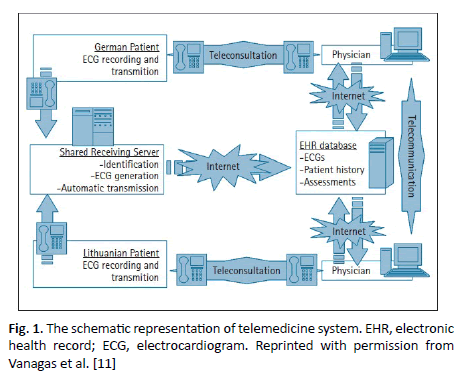
Figure 1. The schematic representation of telemedicine system. EHR, electronic health record; ECG, electrocardiogram. Reprinted with permission from Vanagas et al. [11]
The possibility of ultrasound pictures through real-time wireless transmission was examined in the different studies. For instance, in a study [9], trauma images obtained by the focused assessment with sonography were first hold on an ambulance and then wirelessly transferred to an antenna. In the following, for review at a remote location, trauma images sent through satellite. The results demonstrated that antenna pictures have a decreased quality compared with pictures viewed on-site. In fact, it was observed a 32% declining in the quality of pictures and a 42% reduction in the quality of video clips when they transferred through satellite remotely. However, the improved quality for images was recorded in a research [10]. The authors transmitted echocardiograms made in the field through microwave signal wirelessly to a satellite for viewing offsite. In the comprehensive research, on-site images and formal echocardiography were compared to 12 transmitted studies. The cardiologist reviewers classified good agreement in pericardial effusion (100%), ejection fraction (100%), left ventricle size (92%), and technical quality (83%).
Pre-hospital setting: Ultrasound technique has found a number of applications in pre-hospital setting, such as aeromedical transport [3]. In a study on aeromedical transportation, a number of critical care providers, emergency physicians, technologists, residents, and flight nurses assessed the possibility of ultrasound technique during flight situations. The authors experienced no significant barriers in accomplishing the aeromedical transport. No report on the endangerment of the flights safety was said by pilots [12]. Furthermore, ultrasound technique has the potential to collaborate the in-hospital physicians to get ready for the trauma patients, and to recognize intra-peritoneal bleeding in the field. This technique has been successfully employed in pre-hospital triage setting on victims of an earthquake event who screened by physicians [13]. Despite of the insufficient data, it can be concluded that ultrasound technique prove essential in military situations and disasters for patients triage, as well as the rapid assessment.
In addition, ultrasound can be used as a supplement to the triage System of Simple Triage and Rapid Treatment (START) [14] (Figure 2). In a study [14], clinical measures such as the Glasgow Coma Scale were used to triage patients: expectant (black), immediate (red), delayed (yellow), and ambulatory (green). The charts related to 575 samples of the trauma patients were reviewed. In the following, each subject was appropriated to a START triage category of black, yellow or red.
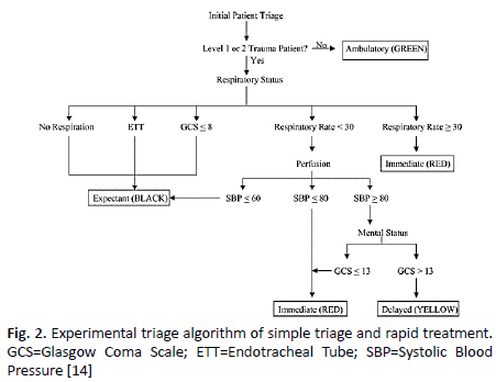
Figure 2. Experimental triage algorithm of simple triage and rapid treatment. GCS=Glasgow Coma Scale; ETT=Endotracheal Tube; SBP=Systolic Blood Pressure [14]
The outcomes of FAST for 360 subjects were indicated that a total of 27 subjects were positive. The researchers detected that 23% of positive FAST tries represent the false positively results, which lead to over triage of yellow subjects to the red class. Moreover, it was observed that 13% of the negative studies were false negatives. It was not observed any benefit for application the FAST assessment as an instrument in order to change triage disposition, albeit it is effortful to depict consequences based on the information provided by the current research [15].
Soft tissue ultrasound: Ultrasound has been utilized with success to check up of injuries of soft tissue in sports medicine and orthopedics. This technique can be employed for evaluation of the soft tissues such as tendons (Figure 3). Ultrasound also can be beneficial particularly when accomplishment a subtle tendon tears assessment [16]. In orthopedic and sports medicine, this technique has been utilized for examination of different situations. For instance, the rotator cuff injuries within the shoulder are the most prevalent injury, which have been assayed through ultrasound [17]. For the focal damages related to rotator cuff tendons, the literature of radiology cites specificities and sensitivities of 94% and 93%, respectively [18]. In addition to assessment of injuries in soft tissue, ultrasound technique has been successfully employed to guide the injections of tendon sheaths, intra-articular and bursal injections, as well as associated aspirations [19]. It is notable that this application of ultrasound is the suitable for surveying of hip injections in sports medicine. For joints such as knee as well as shoulder, it remains to be specified whether ultrasound provides a benefit upon classical procedures that utilize anatomic critical points.
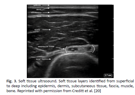
Figure 3. Soft tissue ultrasound. Soft tissue layers identified from superficial to deep including epidermis, dermis, subcutaneous tissue, fascia, muscle, bone. Reprinted with permission from Creditt et al. [20]
Fractures ultrasound: Ultrasound allows the physicians to recognize fractures when customary radiography is not available and or delayed (Figure 4). This application of ultrasound may be employed in the future for preliminary diagnosis of long-bone damage in instable subjects with multi-trauma. It is notably that physicians in emergency department lack enough skill with ultrasound for checking up fractures now [21]. It seems that assessment of fractures by ultrasound is limited in orthopedics because of incapacity to visualize the tissue of medullary, and the highly reflective surface of bone. However, this characteristic provides an opportunity to visualize the intervals of the linear bony cortex easily [22]. In fact, ultrasound can reveal potential fractures that cannot be identified through routine radiography technique. A number of research showed that ultrasound has good and acceptable accuracy clinically in diagnosing occult fractures of the humerus [23], orbital floor [24], femur [25], rib [22], and foot and ankle [26]. For instance, Wang et al. [26] carried out a high-resolution sonography with a 10-MHz lineararray transducer in 270 patients with ankle and foot damages whom were not identified as positive for fracture by using x-ray films. The authors suggested that ultrasound can represent vital information on cortical discontinuities and soft tissue damages in ankle and foot. In fact, ultrasound technique allows emergency physicians to evaluate the process of fracture healing through visualizing the surrounding soft tissue to [27].
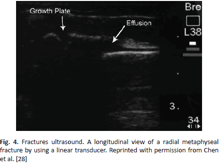
Figure 4. Fractures ultrasound. A longitudinal view of a radial metaphyseal fracture by using a linear transducer. Reprinted with permission from Chen et al. [28]
Ocular ultrasound: Ocular ultrasound is a technique that is employed in the diagnosis of ocular diseases pathology by ophthalmologists [29]. The first application of emergency ultrasound by emergency physicians goes back to 2000 by Blaivas [30], in which the author details two cases of vitreous detachment and globe rupture exactly recognized through emergency ultrasound. Later, several small case series have proposed the application of emergency ultrasound for subjects whose ocular pathology is suspected. Blaivas et al. [30] also examined the precision of emergency ultrasound in ocular pathology 62 subjects specified as retrobulbar hematomas, vitreous hemorrhage, lens dislocation, retinal central artery, globe rupture, vein occlusion, retinal and vitreous detachments, as well as foreign bodies. The researchers employed the computed tomography and ophthalmologic evaluation as the standard reference in their study. They found specificity of 98%, positive predictive value of 97%, sensitivity of 99.9%, and negative predictive value of 99.9%. Furthermore, ocular ultrasound can be used for recognizing of papilledema, which can be a challenging bedside diagnosis in pediatric patients exclusively. Blaivas et al. [31] detected that ocular emergency ultrasound own a specificity of 97%, negative predictive value of 100%, sensitivity of 98%, and positive predictive value of 95%, in 35 subjects with suspected intracranial hemorrhage from aneurysmal hemorrhage or trauma who underwent computed tomography for verification (Figure 5). The current research proposed that ocular ultrasound can be accurately carried out after one hour of hands-on instruction assigned to scanning of ocular pathology by emergency physicians [32]. It remains to be established the capability of ocular emergency ultrasound to affect the decision-making capacity by physicians in emergency department [21].
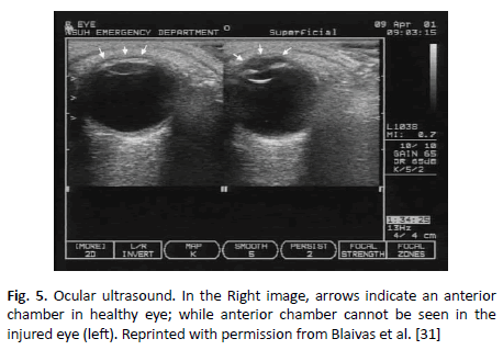
Figure 5. Ocular ultrasound. In the Right image, arrows indicate an anterior chamber in healthy eye; while anterior chamber cannot be seen in the injured eye (left). Reprinted with permission from Blaivas et al. [31]
Paracentesis ultrasound: Literature on the application of real-time ultrasound is limited for evaluate paracentesis. Critical care ultrasound is often performed prior to paracentesis for identification optimal needle insertion point and depth, as well as for recognition ascites (Figure 6) [33]. The major steps of paracentesis in the critical care ultrasound are included: relieving symptoms related to a tense ascites, and then ruleout spontaneous bacterial peritonitis, and finally commence to recognition in novel-onset ascites [33]. In a clinical study [34], the critical care providers used routine means and bedside critical care ultrasound for paracenteses assessment. The authors showed that ultrasound- mediated evaluation of paracentesis had a higher success rate than the non-ultrasound routine means. In fact, the success rate was documented 95% and 61% for diagnose with ultrasound enlightenment and without ultrasound enlightenment, respectively. It is notably, paracentesis in widevolume can be accomplished in the critical care or outpatient setting for those of subjects with chronic ascites, which need the repeated drainage.
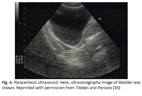
Figure 6. Paracentesis ultrasound. Here, ultrasonography image of bladder was shown. Reprinted with permission from Tibbles and Porcaro [35]
Pneumothorax ultrasound: The findings from research in adult subjects have revealed the critical care ultrasound can be successfully used for discovering the non-traumatic pneumothorax [36]. In a study by Lichtenstein et al. [37], an algorithm for adult subjects was suggested for discovering of occult pneumothorax that not seen on chest radiograph. The results indicated a specificity of 95%, sensitivity of 94%, predictive negative value of 97%, as well as predictive positive value of 73%. This algorithm begins with assessment of lung sliding, and continues with evaluation of A-lines and lung point to find the occult pneumothorax [37]. A number of researches in adult trauma patients have similarly demonstrated the higher accuracy of critical care ultrasound over chest radiograph for discovering pneumothorax in humans [38-40] (Figure 7). The application of ultrasound for detecting pneumothorax in pediatrics remains largely unknown, albeit one case report indicated the detection of pneumothorax by using ultrasound [41].
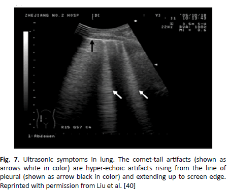
Figure 7. Ultrasonic symptoms in lung. The comet-tail artifacts (shown as arrows white in color) are hyper-echoic artifacts rising from the line of pleural (shown as arrow black in color) and extending up to screen edge. Reprinted with permission from Liu et al. [40]
Foreign bodies' ultrasound: Most extremity wounds have an occult foreign body, which can result in some complications such as inflammation, loss of function, and infection [42]. A backdated study represented that 40% of foreign bodies in the patients arm cannot be recognized in the elementary physical assessment [43]. These missed bodies are considered as the second main reason of claims in the emergency department, as well as they contribute to 6% of all consensuses [42]. Plain radiography technique is used as conventional screening method to detect the foreign bodies in the extremity wounds (Figure 8). Nevertheless, radiopaque objects such as stone, glass, and metal only are able to find in plain radiography, while radiolucent objects such as plastic, wood, and thorns are lost in this technique. So far, a number of imaging procedures have been used in the effort to explain an approach for precise detecting of foreign bodies located in the human soft tissue. For example, Ginsberg et al. [44] compared the ability of different imaging modalities for detection of plastic, glass, and wood. The authors showed that ultrasound detects plastic and wood pieces located between strips of steak submerged in water, while computed tomography, xerography, and plain radiography cannot find these fragments.
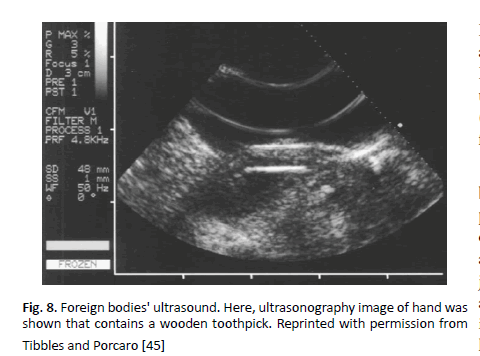
Figure 8. Foreign bodies' ultrasound. Here, ultrasonography image of hand was shown that contains a wooden toothpick. Reprinted with permission from Tibbles and Porcaro [45]
Is the use of bladder ultrasound before the suprapubic aspiration [46] and urethral catheterization [47, 48] in young children. Chen et al., [47] performed a bedside bladder ultrasound in patients younger than two years in front of urethral catheterization (Figure 9). They obtained the sagittal and transverse views of the bladder, and the volume of bladder was measured. The urethral catheterization was postponed until enough level of urine was observed in the study. The results revealed that by using of bladder ultrasound, physician is able to enhance the catheterization success rate from 75% to 95%. Witt et al., [48] similarly observed an enhancement in the success rate of urethral catheterization from 65% to 95%. The authors suggested that the painful repeated methods were avoided in subjects through a non-invasive, fast, and simple method.
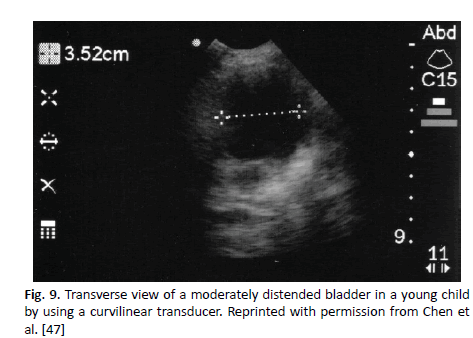
Figure 9. Transverse view of a moderately distended bladder in a young child by using a curvilinear transducer. Reprinted with permission from Chen et al. [47]
Arthrocentesis ultrasound: In the emergency department, swelling and joint pain are typical complaints. Joint effusions are resulted from multiple processes, such as inflammation, connective tissue diseases, infection, and crystal arthopathies [49]. In case of joint effusions, determination a definitive diagnosis often needs synovial fluid analysis [50]. Arthrocentesis of particular joints including ankle and hip can be laborious from a technical perspective, although most joints are be able to tap through the anatomic well-established landmarks. Ultrasound technique localizes the fluid collection and is able to employ in order to guide the needle in the course of the procedure. In ultrasound, a high-frequency probe can visualize fluid as little as one mL [33]. Moreover, ultrasound has a potential to verify the lack of a joint effusion afterward a dry tap.
In contrast to MRI and CT, bedside ultrasound is less expensive and faster to assay the joint effusions in emergency department. Furthermore, ultrasound can be impressively utilized to check up the symptomatic joints- such as hip- with the opposite sides (Figure 10). As an important point, no sedative helper is needed for assessment of patients with low age [49].
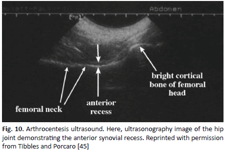
Figure 10. Arthrocentesis ultrasound. Here, ultrasonography image of the hip joint demonstrating the anterior synovial recess. Reprinted with permission from Tibbles and Porcaro [45]
Fortunately, the guidelines for the emergency application of bedside ultrasonography in finding the joint effusions have been precisely reviewed and published. In a review [49], the authors discussed the ultrasound characteristics of joint effusions, and also the specific ultrasound windows utilized for imaging each joint. Among joint effusions, the ankle and hip arthrocenteses are generally considered more laborious in the course of technical issues to carry out. As for hip arthrocenteses, a joint effusion has been diagnosed when a 2 mm discrepancy is appreciated between two sides. Furthermore, the joint effusion has been diagnosed when fluid expanding along the joint capsule measures higher than five millimeters [49]. In a study on a total of 20 hip joint aspirations, which guided by ultrasound, joint fluid was aspirated more quickly when ultrasonography indicated the capsular distance [50]. In a study, Roy et al. [51] reported an effective procedure for ultrasound-guided arthrocentesis of ankle. As for ankle arthrocenteses, an effusion is observed as an anechoic collection in the anterior recess of the talocrural join [51]. It is notably, the presence of fluid is common in the talocrural join, and the clinical conditions and positions would determine whether arthrocentesis is need or not.
Conclusion
Invasive and rapid methods are important to the daily practice of physicians in critical care and emergency departments. Many of these methods, ranging from telemedicine to a simple drainage and incision, can be carried out under emergency and critical care ultrasound guidance, such as telemedicine, pre-hospital setting, soft tissue, fractures, ocular, paracentesis, pneumothorax, bladder, arthrocentesis, and foreign bodies' ultrasound. In fact, emergency medicine and critical care ultrasound has several clinical uses that improve results of patients with life-threatening emergency conditions. Ultrasound imaging improves diagnostic accuracy and provides vital data to emergency physicians to guide management and assist triage subjects to suitable destinations. Time limitations and training requirements are considered as the major challenges to use of ultrasound. The potential for application of this method in the emergency medicine and critical care setting is great; however more studies is needed to provide enough evidence on its clinical impact on patient treating.
Conflict of Interest
None
References
- Soares M, Caruso P, Silva E, Teles JM, Lobo SM, et al. Characteristics and outcomes of patients with cancer requiring admission to intensive care units: a prospective multicenter study. Crit Care Med. 2010;38:9-15.
- Laher AE, McDowall J, Gerber L, Aigbodion SJ, Enyuma COA, et al. The ultrasound 'twinkling artefact' in the diagnosis of urolithiasis: hocus or valuable point-of-care-ultrasound? A systematic review and meta-analysis. Eur J Emerg Med. 2020;27:13-20.
- Bøtker MT, Jacobsen L, Rudolph SS, Knudsen L. The role of point of care ultrasound in prehospital critical care: a systematic review. Scand JTraumaResus. 2018;26:1-14.
- El Sayed MJ, Zaghrini E. Pre-hospital emergency ultrasound: a review of current clinical applications, challenges, and future implications. Emer Med Int. 2013.
- Brenner DJ, Doll R, Goodhead DT, Hall EJ,Land CE, et al. Cancer risks attributable to low doses of ionizing radiation: assessing what we really know. Proc Natl Acad Sci USA. 2003;100:13761-13766.
- Garrett PD, Boyd SY, Bauch TD, Rubal BJ,Bulgrin JR, et al. Feasibility of real-time echocardiographic evaluation during patient transport. J Am Soc Echocardiogr. 2003;16:197-201.
- Urban MJ, Edmondson DA, Aufderheide TP. Prehospital 12-lead ECG diagnostic programs. Emerg Med Clin North Am. 2002;20:825-841.
- Trippi JA, Lee KS, Kopp G, Nelson D,Kovacs R. Emergency echocardiography telemedicine: an efficient method to provide 24-hour consultative echocardiography. J Am Coll Cardiol. 1996;27:1748-1752.
- Strode CA, Rubal BJ, Gerhardt RT, Christopher FL,Bulgrin JR, et al. Satellite and mobile wireless transmission of focused assessment with sonography in trauma. Acad Emerg Med. 2003;10:1411-1414.`
- Huffer LL, Bauch TD, Furgerson JL, Bulgrin J,Boyd SY. Feasibility of remote echocardiography with satellite transmission and real-time interpretation to support medical activities in the austere medical environment. J Am Soc Echocardiogr. 2004;17:670-674.
- Vanagas G, Zaliūnas R, Benetis R, Slapikas R, Smith W. Clinical-technical performance and physician satisfaction with a transnational telephonic ECG system. Telemed J E Health. 2008;14:695-700.
- Price DD, Wilson SR, Murphy TG. Trauma ultrasound feasibility during helicopter transport. Air Med J. 2000;19:144-146.
- Sarkisian AE, Khondkarian RA, Amirbekian NM, Bagdasarian NB, Khojayan RL, et al. Sonographic screening of mass casualties for abdominal and renal injuries following the 1988 Armenian earthquake. J Trauma. 1991;31:247-250.
- Sztanjnkrycer MD, Baez AA, Luke A. FAST ultrasound as an adjunct to triage using the START mass casualty triage system: a preliminary descriptive system. Prehosp Emerg Care. 2006;10:96-102.
- Nelson BP, Chason K. Use of ultrasound by emergency medical services: a review. Int J Emerg Med. 2008;1:253-259
- Jacobson JA. Ultrasound in sports medicine. Radiol Clin N Am. 2002;40:363-386.
- Martinoli C, Bianchi S, Prato N, Pugliese F, Zamorani MP, et al. US of the shoulder: non-rotator cuff disorders. Radiographics. 2003;23:381-401.
- Holsbeek MT, Kolowich PA, Eyler WR, Craig JG, Shirazi KK, et al. US depiction of partial-thickness tear of the rotator cuff. Radiology. 1995;197:443-436.
- Cardinal E, Bureau NJ, Aubin B, Chhem RK. Role of ultrasound in musuloskeletal infections. Radiol Clin N Am. 2001;39:191-201.
- Creditt AB, Joyce M, Tozer J. Skin and soft tissue ultrasound. in: clinical ultrasound. Springer. 2018.
- Legome E, Pancu D. Future applications for emergency ultrasound. Emerg Med Clin N Am. 2004;22:817-827.
- Mariacher-Gehler S, Michel BA. Sonography: a simple way to visualize rib fractures. AJR Am J Roentgenol. 1994;163:1268.
- Patten RM, Mack LA, Wang KY, Lingel J. Nondisplaced fractures of the greater tuberosity of the humerus: sonographic detection. Radiology. 1992;182:201-204.
- Jenkins CN, Thuau H. Ultrasound imaging in assessment of fractures of the orbital floor. Clin Radiol. 1997;52:708-711.
- Watson NA, Ferrier GM. Diagnosis of femoral shaft fracture in pregnancy by ultrasound. J Accid Emerg Med. 1999;16:380-381.
- Wang CL, Shieh JY, Wang TG, Hsieh FJ. Sonographic detection of occult fractures in the foot and ankle. J Clin Ultrascoun. 1999;27:421-425.
- Failla JM, Jacobson J, Van Holsbeeck M. Ultrasound diagnosis and surgical pathology of the torn interosseous membrane in forearm/dislocations. J Hand Surg. 1999;24:257-266.
- Chen L, Moore CL. Bedside ultrasound in the diagnosis and guided reduction of forearm fractures in children. Pediatric Academic Society Annual Meeting. 2005.
- Marin J. Novel applications in pediatric emergency ultrasound. Clin Pediatr Emerg Med. 2011;12:53-64.
- Balivas M. Beside emergency department ultrasonography in the evaluation of ocular pathology. Acad Emerg med. 2000;7:947-950.
- Blaivas M, Theodoro D, Sierzenski PR. A study of bedside ocular ultrasonography in the emergency department. Acad Emerg Med. 2002;9:791-799.
- Blaivas M, Theodoro D, Sierzenski PR. Elevated intracranial pressure detected by bedside emergency ultrasonography of the optic nerve sheath. Acad Emerg Med. 2003;10:376-381.
- Srinivasan S, Cornell TT. Bedside ultrasound in pediatric critical care: A review. Pediatr Crit Care Med. 2011;12:667-674.
- Nazeer SR, Dewbre H, Miller AH. Ultrasound-assisted paracentesis performed by emergency physicians vs the traditional technique: A prospective, randomized study. Am J Emerg Med. 2005;23:363-367.
- Tibbles CD, Porcaro W. Applications of ultrasound. Emerg Med Clin North Am. 2004;22:797-815.
- Lichtenstein DA, Meziere GA. Relevance of lung ultrasound in the diagnosis of acute respiratory failure: The BLUE protocol. Chest. 2008;134:117-125.
- Lichtenstein DA, Meziere G, Lascols N, Biderman P,Courret JP, et al. Ultrasound diagnosis of occult pneumothorax. Crit Care Med. 2005;33:1231-1238.
- Alsalim W, Lewis D. Towards evidence based emergency medicine: Best BETs from the Manchester Royal Infirmary. BET 1: Is ultrasound or chest x ray best for the diagnosis of pneumothorax in the emergency department? Emerg Med J. 2009;26:434-435.
- Jaffer U, McAuley D. Best evidence topic report. Transthoracic ultrasonography to diagnose pneumothorax in trauma. Emerg Med J. 2005;22:504-505.
- Zhang M, Liu ZH, Yang JX, Gan JX,Xu SW, et al. Rapid detection of pneumothorax by ultrasonography in patients with multiple trauma. Crit Care. 2006;10:R112.
- Liu DM, Forkheim K, Rowan K, Mawson JB, Kirkpatrick A, et al. Utilization of ultrasound for the detection of pneumothorax in the neonatal special-care nursery. Pediatr Radiol. 2003;33:880-883.
- Schlager D. Ultrasound detection of foreign bodies and procedure guidance. Emerg Med Clin N Am. 1997;15:895-912.
- Anderson MA, Newmeyer WL III, Kilgore ES Jr. Diagnosis and treatment of retained foreign bodies in the hand. Am J Surg. 1982;144:63-67.
- Ginsburg MJ, Ellis GL, Flom LL. Detection of soft-tissue foreign bodies by plain radiography, xerography, computed tomography, and ultrasonography. Ann Emerg Med. 1990;19:701-703.
- Tibbles CD, Porcaro W. Procedural applications of ultrasound. Emerg Med Clin North Am. 2004;22:797-815.
- Ozkan B, Kaya O, Akdak R, Unal O, Kaya D. Suprapubic bladder aspiration with or without ultrasound guidance. Clin Pediatr. 2000;39:625-626.
- Chen L, Hsiao AL, Moore CL, Dziura JD, Santucci KA. Utility of bedside bladder ultrasound before urethral catheterization in young children. Pediatr. 2005;115:108-111.
- Witt M, Baumann BM, McCans K. Bladder ultrasound increases catheterization success in pediatric patients. Acad Emerg Med. 2005;12:371-374.
- Valley VT, Stahmer SA. Targeted musculoarticular sonography in the detection of joint effusions. Acad Emerg Med. 2001;8:361-367.
- Mayekawa DS, Ralls PW, Kerr RM, Lee KP, Boswell WD. Sonographically guided arthrocentesis of the hip. J Ultrasound Med. 1999;8:665-667.
- Roy S, Dewitz A, Paul I. Ultrasound-assisted ankle arthrocentesis. Am J Emerg Med. 1999;17:300-301


