Case Report - Onkologia i Radioterapia ( 2021) Volume 15, Issue 2
The first report of philadelphia positive chronic myeloid leukemia in an infant presenting merely with leukocytosis (Iran)
Gholamreza Bahoush1*, Babak Abdolkarimi2 and Pourya Salajegheh32Department of Pediatrics, Lorestan University Of Medical Sciences, Lorestan, Iran
3Department of Pediatrics Hematology And Oncology, Children’s Medical Center, Kerman, Iran
Gholamreza Bahoush, Department of Pediatric Hematologist And Oncologist, Faculty Of Medicine, Ali-Asghar Children Hospital, Iran University Of Medical Sciences, Tehran, Iran, Email: gr.bahoushoncologist@gmail.com
Received: 19-Dec-2020 Accepted: 25-Jan-2021 Published: 15-Feb-2021
Abstract
Childhood Chronic Myeloid Leukemia (CML) normally appears in adult group and its occurrence among children and particularly infants is very rare. Adult-Type CML (ATCML), which involves Philadelphia chromosome (Ph1); and commonly, presents with three important findings: huge splenomegaly, hyperleukocytosis and thrombocytosis. In this report, an infant with adult-type CML describes by detail without any common significant findings such as splenomegaly and thrombocytosis. This case is particularly attractive due to its very rare incidence in infancy, and the fact that it is presenting only with leukocytosis.
Keywords
chronic myelogenous leukemia, adult type-cml, Philadelphia chromosome (ph1)-positive cml, leukocytosis
Introduction
Two to 5% of all leukemia cases are observed in pediatric patients. Among the population under 20 years of age, its annual incidence rate is less than one per 100,000. JMML in children below the age of 5 years is observed infrequently. And accordingly, Philadelphia chromosome (Ph1)-positive Chronic Myelogenous Leukemia (CML) or Adult type CML (ATCML) is even rarer in childhood, especially below the age of 5 years. In fact, it is found only in about 3% of the childhood leukemia cases. The Manchester Children’s Tumor Registry has reported only 9 cases of this form in children over the course of 23 years. Sometimes, atypical manifestations are seen in a CML case. An atypical CML with isolated thrombocytosis is different from a typical one in several aspects. In patients of atypical CML, no splenomegaly is noted. In these patients, the number of peripheral leukocyte are normal, and LDH and uric acid levels are lower than those in classic CML, while hemoglobin levels are higher [1]. Regarding the treatment, Imatinib mesylate, a bcr-abl tyrosine kinase inhibitor, is the primary treatment agent for CML in children.
Case Report
A 15-month-old male child presented with no typical manifestations such as fever, hepatosplenomegaly or respiratory distress. Screening tests were conducted on him at 12 months of age for Complete Blood Count (CBC), and White Blood Cell (WBC) was 12000/μl. No significant abnormal finding was observed in the examinations. Physicians recommended a CBC check in two months, which showed a WBC increase to 25000/ μl. Still no abnormality was noted. However, one-month later, his CBC increased to 32000/μl, and he was referred to our center for further evaluations.
During a routine full blood count test, surprising observations were made. Following that, further physical examinations and lab tests, shown in Table 1, yielded unremarkable results [2].
Tab. 1. Patient’s paraclinic test
| Variable | value | Definition |
|---|---|---|
| Hb (g/dL) | 9.8 | |
| WBC/µL | 32000 | Lymphosyte: 23%, Neutrophil: 50% Band: 5%, Basophil: 8%, Eosinophil: 5% Metamyelocytes: 4%, Myelocyte: 5% |
| Platelet/µL | 341000 | |
| ALT | 15 | |
| AST | 38 | |
| LDH | 794 | |
| T.Bil | 0.5 | |
| D.Bil | 0.1 | |
| BUN | 8 | |
| Creat | 0.5 | |
| CRP | 2 | Normal Values: less than 3 mg/l |
| ESR | 20 | |
| G6PD | sufficint | |
| TORCH study | ||
| Hb F | 18.50% | |
| Hb A1 | 80% | |
| Hb A2 | 2.50% | |
| Blood culture | negative | |
| Immunoelectrophoresis | normal | |
| ABL-BCR ratio | 0.50% | |
| Mean copy for control ABL-BCR gen | 79125 | |
| Mean copy for fusion ABL-BCR gen | 422 | |
| Immunoelectrophoresis | normal |
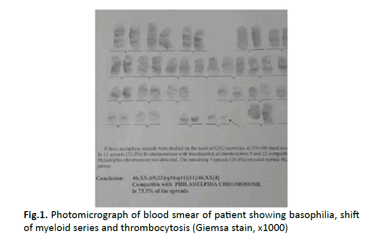
In the peripheral blood smear, microcytic hypochromic erythrocytes with many dacrocytes, polychromatophilic Red Blood Cells (RBCs), and normoblasts were observed. The number of leukocyte was 90,600 and the differential count was as 53% neutrophil, 14% myelocyte, 2% metamyelocyte, 16% basophil, 17% eosinophil, 25% lymphocyte. Bone marrow aspiration results showed increased abnormal megakaryocytes and bone marrow fibrosis. Monolobated and multinucleated megakaryocytes with hyperchromatic and pleomorphic nuclei were seen as well. The neutrophils showed dysplastic features such as hypo-, hypersegmentation, and hypogranulation. Many giant platelets and platelet aggregates were identified. Individual platelets showed noticeable variations in size. ABLBCR quantitative RT-PCR report, demonstrated a high copy number (more than 100%) and 46 XX with the presence of the Philadelphia chromosome and additional cytogenetic abnormalities (q34 ؛ q11) (Figure 1). Consequently, a diagnosis of a Ph1-positive type of CML was made and a treatment regimen started with original Imatinib Mesylate.

Figure 1: Photomicrograph of blood smear of patient showing basophilia, shift of myeloid series and thrombocytosis (Giemsa stain, x1000)
Discussion
Infantile Philadelphia chromosome (Ph1)-positive CML is extremely rare. Pediatric CML is a type of chronic myeloid leukemia occurring in infants and children. Unusually, it occurs in infants less than 6 months of age. The incidence of CML increases with age and no effective ethnic or genetic factor has been identified in this disease. However, ionizing radiation proved to be a risk factor for this disease. Since CML is greatly infrequent, conducting hematologic studies for gaining a deeper understanding is difficult.
Philadelphia Chromosome (Ph (1))-positive Chronic Myelogenous Leukemia (CML) in a child below the age of 3 is very unthinkable. There are a few cases including one reported regarding a 3-year-old male child, however, our patient is 15 months. 3 Hugo Castro-Malaspina reported the diagnostic characteristics of 39 children with Philadelphia chromosomepositive chronic myelocytic leukemia (Ph1-positive CML) [3], during 1963 to 1976 in the Hôpital Saint-Louis of Paris. Thus, our patient seems to be one of the youngest case of this disease. In conclusion, this study suggests that Ph1-positive CML of childhood exhibits the same course, incidence of blastic crisis, and survival as it does in adults. It also indicates that treatment regimen with moderate chemotherapy, such as busulfan, does not affect the duration of survival. Therefore, newly found therapeutic approaches are urgently needed for treating this disorder in children.
Frédéric Millot reported (2005) the clinical and biological characteristics of diagnosis in children and adolescents with Chronic Myelogenous Leukemia (CML). This is the most extensive reported series of CML diagnosed in children and adolescents. This study theorizes that the characteristics of CML seem to differ in children compared with previously published reports on adult cases. For instance, particularly, the leukocyte counts are often higher in children. Symptoms were more common in patients with splenic enlargement, which was observed in 70% of the cases, and higher leukocyte counts.
Considerable increased leukocyte counts were common as well (median white blood cell count: 242 × 109/L). Our patient was a case of leukocytosis without splenomegaly [4]. In 1981 reported two CML cases and a review of 55 cases. Similar to the present case, five patients of his cases did not have splenomegaly during the whole course. However, none of the patients were infants at the time of diagnosis. That was a rare feature in his CML patients [1,5]. Studied CML cases with isolated thrombocytosis. No splenomegaly was seen in these patients, peripheral leukocyte numbers and formulae were normal, LDH and uric acid levels were lower than in classic CML, while haemoglobin levels were higher.
More than 30% of megakaryocytes in the chronic stage of CML are smaller than normal, with hypolubulated nuclei. Nonetheless, there are instances of Philadelphia chromosomepositive (Ph+) CML with isolated thrombocytosis (CML-T) or slightly elevated granulocyte numbers. This special form of CML is described as Essential thrombocythemia, and is more prevalent in women. It is characterized by the absence of splenomegaly, and smaller megakaryocytes with hypolobulated nuclei in the bone marrow [1].
Reema Al Hayek reported the first case of an infantile CML in accelerated phase demonstrating three-way Ph chromosome variant t (9; 22; 14) (q34; q11.2; q32). The authors concluded that it is important to realize that chromosomal abnormalities including variant Ph chromosome appearing as a new cytogenetic abnormality in addition to the classical Ph (clonal evolution) has a bad prognostic value [6].
Generally speaking, children tend to exhibit juvenile myelomonocytic leukemia, which is a type of chronic leukemia with monocytosis and no involvement of Philadelphia chromosome. However, in the present study, the patient did not meet any criteria for JMML including absolute monocyte count >1000/μL, <20% blasts in the bone marrow, absence of the BCR-ABL fusion gene, and mandatory genetic abnormality such as somatic mutation in KRAS, NRAS, or PTPN11 , monosomy 7, NF1 gene mutation, germline CBL mutation, and loss of heterozygosity of CBL [7].
Transient Abnormal Myelopoiesis (TAM) that can develop in newborns who have Down syndrome is the most prominent Philadelphia negative differential diagnosis in myeloid disorder. It usually fades needing no intervention within the first 3 months of life. Infants who have TAM have an increased chance of developing AML before the age of 3. TAM is also known under the names transient myeloproliferative disorder or transient leukemia. GATA1 mutation and Ph-chromosome are differentiating laboratory tests for TAM and Infantile Philadelphia chromosome (Ph1)-positive CML. The patient in this report did not have any of these genetic abnormalities [8]. The age of 3. TAM is also known under the names transient myeloproliferative disorder or transient leukemia. GATA1 mutation and Ph-chromosome are differentiating laboratory tests for TAM and Infantile Philadelphia chromosome (Ph1)- positive CML. The patient in this report did not have any of these genetic abnormalities [8].
Conclusion
Ph-positive CML in a child below the age of 5 is extremely rare yet possible. The absence of splenomegaly was another unexpected finding in this CML patient. CML can emerge with an atypical manifestation in infants, which causes delayed diagnosis. In this paper, we reported the first case of infantile CML presented merely with isolated leukocytosis. Therefore, CML should be considered as a possible scenario in all cases of isolated leukocytosis, even in the absence of splenomegaly.
Ethics Approval
Institutional review board approval for the case report is not required at our institution. Following the ethical principles, names of the patient was not pointed in the paper and the rights of the subject were protected. The patient received treatment consistently with the current standard of care
Consent for Publication
Written informed consent was obtained from the parents of the patient for publication of this case report and any accompanying images.
Acknowledgement
The authors thank the Rasoul Akram Hospital Clinical Research Development Center (RCRDC) for its technical and editorial assists.
Conflict of Interest
The author reports no conflicts of interest in this work.
References
- Yilmaz M, Guvercin B, Konca C. Cases of chronic myeloid leukemia presenting with isolated thrombocytosis: case reports and review of the literature. Int J Clin Exp Med. 2016;9:8799-8802.
- Bahoush G, Alebouyeh M, Vossough P. Imatinib Mesylate (Glivec) in Pediatric Chronic Myelogenous Leukemia. IJHOSCR. 2009;3:8-13.
- Mishra A, Tripathi K, Mohanty L, Pujari S. Philadelphia Chromosome Positive Chronic Myelogenous Leukemia in A Child: A Case Report. Indian J Hematol Blood Transfus. 2010;26:109–110.
- Arashi K, Aibiki T, Higo O. Chronic Myelogenous Leukemia without Splenomegaly: A Report of 2 Cases and a Review of 55 Cases in Literature. J-stage J [Article in Japanese]. 1981;22:159-177.
- Malaspina HC, Schaison G, Briere J. Philadelphia Chromosome-Positive Chronic Myelocytic Leukemia In Children Survival And Prognostic Factors. Cancer. 1983;52:721-727.
- Hayek RA, Omar H, Dayel AA, Raslan H, Dridi W, et al. The First Case Report of an Infant with Three Way Philadelphia Chromosome Variant T (9; 22; 14) (Q34; Q11.2; Q32) Chronic Myeloid Leukemia, King Fahd Specialist Hospital Dammam Experience, Kingdom of Saudi Arabia. Canc Therapy Oncol Int J. 2017;4:1-4.
- Chang YH, Jou ShT, Lin DT, Lu MY, Lin KH. Differentiating Juvenile Myelomonocytic Leukemia From Chronic Myeloid Leukemia in Childhood. J Pediatr Hematol/Oncology. 2004;26:236-242.
- Zipursky A. Transient leukaemia--a benign form of leukaemia in newborn infants with trisomy 21. Br J Haematol. 2003;120:930-938.


