Case Report - Onkologia i Radioterapia ( 2023) Volume 17, Issue 8
Retroperitoneal schwannoma as a rare cause of abdominal pain in a patient with ulcerative colitis:A case report
Farshad ShouhaniMehdi Eshaghzadeh, Department of Radiology, Taleghani Hospital, Shahid Beheshti University of Medical Sciences, Tehran, Iran, Email: mehdie68@gmail.com
Received: 24-Jul-2023, Manuscript No. OAR-23-109110; Accepted: 23-Aug-2023, Pre QC No. OAR-23-109110(PQ); Editor assigned: 31-Jul-2023, Pre QC No. OAR-23-109110(PQ); Reviewed: 05-Aug-2023, QC No. OAR-23-109110(Q); Revised: 21-Aug-2023, Manuscript No. OAR-23-109110(R); Published: 29-Aug-2023, DOI: -
Abstract
Retroperitoneal schwannomas are extremely rare tumors that originate from Schwann cells around the peripheral nerves. The symptoms are nonspecific and there is no pathognomonic radiologic feature for preoperative diagnosis. Complete resection and histopathologic examination are the gold standard for diagnosis and treatment. Here, we present a 54 years old male with a history of abdominal pain and a 47*33 mm abdominal mass at CT scan that diagnosed as schwannoma after a percutaneous ultrasound-guided biopsy and microscopic examination was performed.
Keywords
retroperitoneal schwannoma, abdominal pain, ulcerative colitis
Introduction
Schwannomas or neurilemmomas are rare tumors that originate from the Schwann cells around the peripheral nerves. These tumors are often benign but malignant types may be associated with Recklinghausen disease [1].
Schwannomas occur around the peripheral nerves mostly in the head and neck region. Retroperitoneal schwannomas are rare and comprise only about 0.7% of these tumors [1, 2]. The symptoms of retroperitoneal schwannomas are nonspecific and the main symptoms are abdominal distention and dull abdominal pain [3].
Histologically they are encapsulated tumors, originate from neural crest cells and are composed of spindle cells organized in Antoni A (high cellularity) and Antoni B (low cellularity) regions [1,2]. In immunohistochemistry, schwannomas are diffusely positive for S100 protein in the cytoplasm of the tumor cells [4].
We present here a case of benign retroperitoneal schwannoma in a patient with ulcerative colitis.
Case Presentation
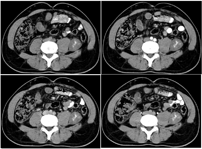
A 54-years-old man with a 10 years history of ulcerative colitis and 2 years history of primary sclerosing cholangitis presented with vague, predominantly left lower quadrant abdominal pain that was present for 3 months. The patient was under treatment with mesalamine. Laboratory test results were unremarkable. Ultrasonography showed a heterogenous mass measuring about 5 cm in left lower quadrant. Computed Tomography (CT) scan with and without IV contrast injection was performed and revealed a 47 mm×33 mm heterogenous predominantly hypodense mass with central area of hyperdensity (intratumoral hemorrhage or calcification) in left retroperitoneal space between left psoas and left iliacus muscles without significant contrast enhancement (Figure 1).

Figure 1: A well-defined predominantly hypodense mass with a central hyperdense area is seen adjacent to the left psoas muscle in non-enhanced CT scan (A). No contrast enhancement is seen in arterial (B), Portal (C), and delayed phase of contrast-enhanced CT scan. Biopsy confirmed the diagnosis of schwannoma.
Ultrasound-guided biopsy performed and no complication occurred after the procedure. On microscopic examination, the mass composed of mostly hypocellular fascicles of bland spindle cells with foci of nuclear palisading. On immunohistochemistry study, the spindle cells were diffusely and strongly positive for S100 protein and negative for SMA, Desmin and CD34. These finding was compatible with schwannoma and the patient candidate for surgery
Discussion
Schwannomas are soft tissue tumors derived from the Schwann cells of peripheral nerve sheaths mostly in the neck and limb regions and they found rarely in retroperitoneum [5]. Retroperitoneal Schwannoma mostly affects patients aged 20 to 50, with a female predominance [6].
Schwannomas are typically benign tumors that rarely progress to malignancy unless they are associated with von Recklinghausen's disease [1].
In retroperitoneal region, RPSs are usually clinically silent and asymptomatic until they gain sufficient size and cause compressive symptoms including vague abdominal/low back pain or abdominal distention. Benign schwannomas do not invade to adjacent organs. They also frequently recognized incidentally on imaging studies performed for other reasons [6].
Preoperative diagnosis of retroperitoneal schwannoma is often difficult. There are no characteristic ultrasound, CT or MRI features to differentiate schwannoma from other differential diagnoses such as paraganglioma, pheochromocytoma, fibrosarcoma, liposarcoma and ganglioneuroma [5].
Ultrasonography can be helpful for detecting this tumor and may show well-defined retroperitoneal hypoechoic heterogenous mass [7]. CT scan reveals low or mixed attenuation mass with welldefined borders. Degenerative changes such as cystic degeneration, calcification, hemorrhage and hyalinization may occur in RPSs and contribute to heterogeneity of these tumors in CT scans [8]. On MRI images, schwannoma is typically hypointense on T1- weighted images and hyperintense on T2-weighted images [5]. Since MRI allows for better visualization of the tumor's origin, vascular architecture, and involvement of other organs, it can be used to image retroperitoneal tumors [1].
CT-guided core-needle biopsy and fine-needle aspiration are helpful and reliable for diagnosis of retroperitoneal schwannoma if the sample contains enough Schwann cells for microscopic examination but because of the risk of hemorrhage, infection and tumor seeding many clinicians do not recommend preoperative image-guided biopsy for diagnosis [1].
Because imaging studies and percutaneous biopsy have limitations in correct preoperative diagnosis, complete surgical resection is the standard treatment and recommended in almost all cases and provides the precise pathological examination [9].
On gross inspection, retroperitoneal schwannomas are solitary, well-defined and firm tumors. Microscopically they consist of spindle cells in areas with high (Antoni A) and low (Antoni B) cellularity [1, 2].
Malignant transformation of retroperitoneal schwannoma and also recurrence after surgical excision has been reported but the prognosis of these tumors is excellent. Close follow up after surgical resection is suggested [1, 9].
Conclusion
Retroperitoneal schwannoma is a rare and usually benign tumor without specific symptoms or characteristic radiologic features. Imaging-guided biopsy can be helpful in diagnosis but complications may occur. Preoperative diagnosis is difficult and complete surgical resection is the treatment of choice. The prognosis is good and recurrence is rare but requires long-term follow up by CT scan.
References
- Caliskan S, Gumrukcu G, Kaya C. Retroperitoneal Ancient Schwannoma: A Case Report. Rev Urol. 2015;17:190-193. [Google Scholar] [Crossref]
- Wollin DA, Sivarajan G, Shukla P, Melamed J, Huang WC,et al. Juxta-adrenal Ancient Schwannoma: A Rare Retroperitoneal Tumor. Rev Urol. 2015;17:97-101. [Google Scholar] [Crossref]
- Li Q, Gao C, Juzi JT, Hao X. Analysis of 82 cases of retroperitoneal schwannoma. ANZ J Surg. 2007;77:237-40. [Google Scholar] [Crossref]
- Souza LCA, Pinto TDA, Cavalcanti HOF, Rezende AR, Nicoletti ALA, et al. Esophageal schwannoma: Case report and epidemiological, clinical, surgical and immunopathological analysis. Int J Surg Case Rep. 2019;55:69-75. [Google Scholar] [Crossref]
- Harhar M, Ramdani A, Bouhout T, Serji B, El Harroudi T. Retroperitoneal Schwannoma: Two Rare Case Reports. Cureus. 2021;13:e13456. [Google Scholar] |[Crossref]
- Safwate R, Wichou EM, Allali S, Dakir M, Debbagh A, et al. Retroperitoneal Schwannoma: Case report. Urol Case Rep. 2021;35:101519. [Google Scholar] | [Crossref]
- Lopes CV, Zereu M, Furian RD, Furian BC, Remonti TA. Retroperitoneal schwannoma diagnosed by endoscopic ultrasound-guided fine-needle aspiration. Endoscopy. 2014;46 Suppl 1 UCTN:E287-8. [Google Scholar] [Crossref]
- Cury J, Coelho RF, Srougi M. Retroperitoneal schwannoma: case series and literature review. Clinics (Sao Paulo). 2007;62:359-62. [Google Scholar] [Crossref]
- Wu Q, Liu B, Lu J, Chang H. Clinical Characteristics and Treatment Strategy of Retroperitoneal Schwannoma Adjacent to Important Abdominal Vessels: Three Case Reports and Literature Review. Front Surg. 2020;7:605867. [Google Scholar] [Crossref]


