Research Article - Onkologia i Radioterapia ( 2023) Volume 17, Issue 7
Morphometric evaluation of parenchymal and stromal constituents of the apparently normal liver tissue adjacent to adenocarcinomatous pathology
Maher Finjan Taher1*, Munqith Mazin Mghamis1, Anwar Madlool Al-Janabi2 and Salih Mahdi Al-Khafaji12University of Kufa, College of Medicine, Department of Biochemistry, Iraq
Maher Finjan Taher, University of Kufa, College of Medicine, Department of Anatomy, Iraq, Email: munqithm.almahmood@uokufa.edu.iq
Received: 15-Apr-2023, Manuscript No. OAR-23-97642; Accepted: 04-Jul-2023, Pre QC No. OAR-23-97642 (PQ); Editor assigned: 08-May-2023, Pre QC No. OAR-23-97642 (PQ); Reviewed: 25-Jun-2023, QC No. OAR-23-97642 (Q); Revised: 02-Jul-2023, Manuscript No. OAR-23-97642 (R); Published: 09-Jul-2023
Abstract
Adenocarcinoma of liver from a primary site is common. This study was formulated for rising the attentions about validity of the apparently normal tissue histology adjacent to cancerous tissue for investigating risk factors that could be associated with or leading to cancers. This issue was investigated by this retrospective study done on slides of adenocarcinoma of liver that were obtained from hospital of Al-Sadder in Najaf city, morphometric parameters including; the number of binucleated/ mononucleated hepatocytes, evaluation of the stromal, parenchymatous, vascular and biliary tissue constituents. The statistical analysis provides conclusive data that could be valuable database for researchers working in the field of histochemical changes associated with cancer.
Keywords
Morphometry, Liver adenocarcinoma
Introduction
The worldwide problem of liver cancer is extensive. Conferring to 2020 evaluations, liver cancer is the sixth greatest frequently detected cancer and the third greatest frequent reason of cancer death [1]. The primary malignant liver tumors arise from; the hepatocytes giving rise to hepatocellular carcinoma, those arise from biliary epithelial cells form cholangiocarcinoma and biliary cystadenocarcinoma), while endothelial cells derived tumours are called angiosarcoma or epithelioid hemangioendothelioma), the combinations of these cells with mesenchymal cells form hepatoblastoma [2]. The hepatocellular-cholangiocellular carcinoma represent two different tumors. Secondary liver tumors that metastasized from a primary site are mostly malignant [3].
Adenocarcinoma means cancer in the glands that line many body tissues, involving prostate, breast, esophagus, pancreas, stomach, colon, rectum, and lungs. Spreading adenocarcinoma in the liver from undiagnosed primary spot is common. The histologic picture of this pathology usually is not helpful because it simulates that of primary localizations [4,5].
Many debates were questioned about the apparently normal tissue histology adjacent to cancerous tissue in regard to the validity of considering this tissue as a normal in regard to the histological and histochemical aspects [6].
Former researcher described variable morphometric parameters of the normal liver histology; these parameters were considered as objective criteria of normal liver function. These parameters described include; the hepatocyte's nuclear size, the frequencies of di or tetra nuclei, and was concluded that the incidence of nuclear polyploidy increased with growing age [7]. Morphometric characteristics of perisinusoidal cells of liver tissue was also described in previous articles. Morphometric evaluation of liver tissue not only described for human, but also described for laboratory animals [8].
This study aims to evaluate the morphometric parameter of parenchymal and stromal constituents in the apparently normal liver tissue surrounding adenocarcinomtous pathological changes.
Materials and Methods
In this retrospective study, the total number of slides of adenocarcinoma liver biopsy examined was fifteen slides. The se slides were obtained from hospital of Al-Sadder in Najaf city,some of the slide that did not contain substantial apparently normal liver tissue were excluded.
The histological tissue preparation of these slide had been done as part of the routine investigation for the patient according to the procedure described in the text book [9] (Bancroft histochemistry).
The paraffin blocks collected were sectioned using a manual rotary microtome (Ziess, Germany), sections of 4 μm–5 μm thickness were obtained. Mounting of these sections were done on glass slides and hematoxylin and eosin staining was routinely accomplished.
Calculation of the number of binucleated/ mononucleated hepatocytes was done by consuming the grid of eye piece microscopic morphometry, and counting of the number of dots in the grid at the single and dual hepatocytes. The samples calculated and examined in arbitrarily nominated high power fields, the numbers found were converted as a fraction [10].
The evaluation of the stromal, parenchymatous, vascular and biliary tissue constituents consuming identical microscopic grid, the number of points at the stromal (septa, hepatic tracts), parenchymal (liver cells), vascular (capillary sinusoids, portal blood vessels, central veins) and biliary system (bile ducts) of the apparently normal liver tissue were calculated. Also, samples studied were examined in arbitrarily nominated high power fields, and the numbers obtained were converted as a fraction [10].
The statistical processing of the results was performed using the Microsoft Excel and SPSS software. The resulting calculated mean and the average standard error were used for statistical evaluation.
Results
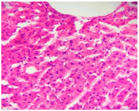
In this study, the morphometric parameters of stromalparenchymatous component of the apparently normal morphological liver tissue adjacent the liver tissue with features of adenocarcinoma maintained the radial arrangement of liver cells around the central vein, and showed apparently normal sinusoids and portal triads (Figure 1).

Figure 1: Apparently normal liver tissue around the adenocarcinomatous pathology H&E. X400.
The morphometric parameters of hepatocytes were found to be as following:
1. The average quantity of mononuclear hepatocytes was 91.25% ± 6.5%.
2. The average quantity of binucleated hepatocytes was 7.5% ± 1.6%.
3. Hepatocytes with fat vacuoles 0.3% ± 0.1%
The stromal-parenchymal percentages were found to be as following:
1. Parenchyma 77.5% ± 4.3%.
2. Liver stromal tissue (counting blood vessels and bile ducts) 22.5% ± 2.6%.
The morphometric evaluation of the liver tissue was as following:
1. The hepatocytes 77.5% ± 4.3%.
2. The portal tracts 2.7% ± 0.6%.
3. The central veins 8.1% ± 1.4%.
4. The sinusoids 9.2% ± 1.3%.
5. The bile ducts 2.5% ± 0.2%.
Discussion
The liver tissue is not homogeneous; it shows variability with characteristic geometry [10]. This research affords a statistical analysis of apparently normal liver tissue of adult human in cases of adenocarcinoma. It was documented in the results of this study that recognizing the histological assessment of apparently liver tissue could be evaluated quantitatively. The current study provides a database on which to make quantitative and qualitative statements about this tissue. The documentation of the normal criteria of this tissue could contribute in upcoming studies of the liver response to injury. Also, the results offer quantitative provision for a pathologist’s report to the physician accomplishment of the biopsy.
The literatures published did not discuss any of the data regarding morphometric parameters of the stromal-parenchymal components of the apparently normal liver tissue around adenocarcinomatous masses. The resulting morphometric data stromal-parenchymatous component reported by this study can be used to be compared with a control group in a study that may have a facility for obtaining normal liver biopsy may be from autopsy. The biopsy of liver is regarded as the best method for assessing the normality of liver tissue, however, this procedure is invasive. It is difficult to obtain normal liver biopsy from a volunteer [11, 12]. If such a comparison is performed, the answer to the question; could research work on the apparently normal liver tissue surrounding a pathology in the liver been considered normal. At least from histological point of view. Accordingly, histochemical techniques for staining of this apparently normal liver tissue may be considered as a valid tool to investigate predisposing factors leading to a specific pathology [13].
Conclusion
The statistical analysis of this morphometric study of the ratio of stromal/parenchymal constituent of the apparently normal liver tissue surrounding adeno carcinomatous pathology opens a new line of research work that is directed for answering to the question; could research work on that tissue been considered normal and it be involved for histochemical staining to investigate predisposing factors leading to a specific pathology.
Acknowledgment
Special thanks to the department of Human Anatomy in the College of Medicine/ Alnahrain University especially to Prof. Dr. Hayder Jawad Kadhim, and great appreciation to the unit of scientific research and cancer study at University of Kufa particularly to Prof. Dr. Rihab Almudhaffer.
References
- Sung H, Ferlay J, Siegel R, Laversanne M, Soerjomataram I. Global cancer statistics 2020: GLOBOCAN estimates of incidence and mortality worldwide for 36 cancers in 185 countries. CA Cancer J Clin. 2021.
- Goodman ZD. Neoplasms of the liver. Mod Pathol. 2007;20: S49-60.
- Quaglia A. Hepatocellular carcinoma: a review of diagnostic challenges for the pathologist. J Hepato Cell Carcinoma 2018; 5:99.
- Ahmed F, Mustafa EM, Basim Sh. Molecular biology of normal mammary tissue surrounding breast cancer of Iraqi women through genetically fluorescent in situ hybridization of ERBB2, TOP2A and C-MYC in relation to estrogen and progesterone. Thesis Submit. Coll. Med. –Al-Mustansirya Univ. (2018).
- Tot T. Adenocarcinomas metastatic to the liver: the value of cytokeratins 20 and 7 in the search for unknown primary tumors. Cancer: Interdiscip. Int. J. Am. Cancer Soc. 1999; 85:171-7.
- Miyama Y, Fujii T, Murase K, Takaya H, Kondo F. Hepatoid adenocarcinoma of the lung mimicking metastatic hepatocellular carcinoma. Autopsy Case Rep. 2020;10.
- Ranek L, Keiding N, Jensen ST. A morphometric study of normal human liver cell nuclei. Acta Pathol Microbiol Scand Pathol.1975; 83:467-76.
- Sztark F, Dubroca J, Latry P, Quinton A, Balabaud C et al. Perisinusoidal cells in patients with normal liver histology: a morphometric study. J Hepatol., 1986; 2:358-69.
- Junatas KL, Tonar Z, Kubíková T, Liška V, Pálek R, et al. Stereological analysis of size and density of hepatocytes in the porcine liver. J Anat. 2017; 230:575-88.
- Suvarna KS, Layton C, Bancroft JD. Bancroft's theory and practice of histological techniques E-Book. Elsevier health sci.; 2018.
- Kokha MK, Landini G, and Iannaccone PM. Fractal geometry in rat chimeras demonstrates that a repetitive cell division program may generate liver parenchyma. Dev Biol 1994; 165:545-555.
- Ludwig J, Wiesner RH, LaRusso NF. Idiopathic adulthood ductopenia: a cause of chronic cholestatic liver disease and biliary cirrhosis. J. Hepatol. 1988; 7:193-9.
- Neuberger J, Patel J, Caldwell H, Davies S, Hebditch V, et al Guidelines on the use of liver biopsy in clinical practice from the British Society of Gastroenterology, the Royal College of Radiologists and the Royal College of Pathology. Gut. 2020; 69:1382-403.


