Case Report - Onkologia i Radioterapia ( 2022) Volume 16, Issue 11
Malignant phyllodes tumour with lymph node metastasis
Oula Fouad Hameed1* and Zainab Abdulkareem Maktoof1Oula Fouad Hameed, Department of Oral Diagnosis, College of Dentistry, University of Basrah, Iraq, Email: medicalresearch64@yahoo.com
Received: 21-Oct-2022, Manuscript No. OAR-22-77937; Accepted: 20-Nov-2022, Pre QC No. OAR-22-77937 (PQ); Editor assigned: 23-Oct-2022, Pre QC No. OAR-22-77937 (PQ); Reviewed: 06-Nov-2022, QC No. OAR-22-77937 (Q); Revised: 09-Nov-2022, Manuscript No. OAR-22-77937 (R); Published: 28-Nov-2022, DOI: 0
Abstract
A 35-year-old woman presented with a left breast mass which was suspected to be a fibroadenoma, excision was done. The tumor size was approximately 6 cm. The histopathological diagnosis was borderline phyllodes tumor. After that, the tumor re-cure 2 times within a short period and the last rapid recurrence within 3 months from the last excision and reaching a large size of 8 cm infiltrating most of the breast. In ultrasonography, there was a swelling of the left axillary lymph nodes. So, mastectomy with axillary lymph nodes excision was performed. The malignant nature of the tumor and the metastasis to the lymph node were confirmed by the pathological examination of the specimens. A few days later a chest CT scan revealed pulmonary nodules. Thus, in this report, we present a case of a malignant phyllodes tumor with an extremely rare lymph node metastasis, which shows later pulmonary metastasis and is now patient on treatment.
Keywords
breast tumor, phyllodes, lymph node, metastasis, malignant.
Introduction
Phyllodes Tumors (PTs) of the breast are rare fibro epithelial tumors seen in the breast, they can be of a benign behaviour that mimics fibro adenoma but with a chance of recurrence if wide margins are not excised and some of these tumors may present borderline features or with malignant pathological features that can metastasize distantly [1]. This tumor forms a leaf-like structure, and its size measures about 4 cm but previous reports mentioned a larger size of 10 cm or more, it accounts for 0.3% – 1 % of all tumors of the breast and about 10%-15% of it are considered malignant with a percentage of 10%-26 % are metastasize [2].
Phyllodes tumors mainly occur in the 4th to 6th decades of life but they also could see in younger age patients who presented with a complaint of a palpable mass in the breast with a distortion of the breast architecture in a mammogram. These tumors grow in a horizontal-radial pattern, also the rate of growth may lead to skin ulceration and hemorrhage with an increased risk of infection, however, there is an elevated risk of metastasis to lungs and bones [3].
Axillary lymph node metastases are rare so most authors have concluded that removal of axillary lymph nodes is not warranted unless pathologically involved [4].
In this report, we present a case of malignant phyllodes tumor with lymph nodes metastasis.
Case Presentation
We reported a case of a 36-year-old female who presented to Al-Mawadda Specialized Surgical Hospital/Basrah city with a recurrent breast mass. At presentation patient's medical history included a surgical excision of a left breast borderline phyllodes tumor (8 months prior) and no history of any type of cancer in her family.
On clinical examination, show big recurrent tumor masses were found in the left breast. There was no pain and the overlying skin is normal.
The US examination described a large, well-defined mu ltiple regular outline hypoechoic lesion in different sizes and sites giving a feature of a large benign breast lesion with inflammatory breast parenchyma of the previous operation. This was re-cure (2 times) within a short period (about 2 and half months’ period)
with a size (6/7 cm) and the surgical treatment each time was tumor excision with breast-conserving, in each time the diagnosis was borderline phyllodes tumor and margin cannot be assessed because the specimens were fragmented. Then the tumor re-cures within about (3 months later) and enlarged considerably in the previous (3 months) from the last excision and reaches a large size of about (8 cm) occupying most of the breast.
The U/S shows an ill-defined mass lesion in the left breast with a round enhanced lymph node at a left axillary area (malignant lymph nodes). Surgical treatment Mastectomy with axillary lymph nodes excision performed.
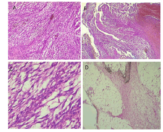
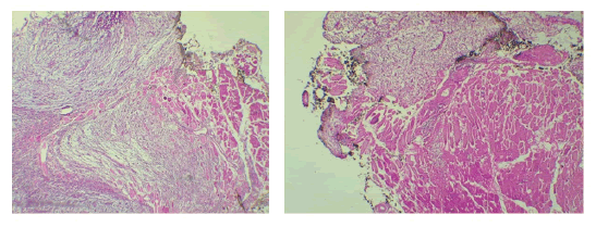
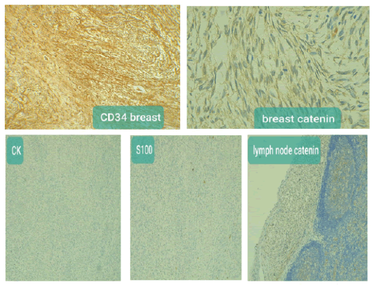
The final pathology examination showed a malignant phyllodes tumor of the left breast (Figure 1), with axillary lymph node metastasis (Figure 2), and involvement of surgical resection margins by the tumor (Figure 3). The immunohistochemistry (IHC) study confirmed malignant PT (Figure 4). CD34: Positive in tumor cells. Negative (Ck, B. catenin, and S100).
Figure 1: A) Moderate-to-marked stromal hyper cellularity with moderateto-marked atypia (B) Stromal overgrowth (H&E, 4) with scarce epithelial elements. (C) Increased mitotic figures (H&E, 40). (D) Infiltration into the surrounding adipocytes (H&E, 4).
Figure 2: Axillary lymph node metastasis.
Figure 3: Involvement of surgical resection margins by tumor.
Figure 4:Immunohistochemistry (IHC) confirmed malignant PT (CD34: Positive in tumor cells. Negative (Ck, B. catenin and S100))
Then the patient did a CT scan of the chest and upper abdomen (native and contrast study), showing multiple iso-dense soft tissue masses lesion largest in the right upper lobe hilar in a location about (45 mm × 50 mm) enhanced after contrast study with mild right pleural effusion with other masses in left lung round in shape features of secondary lung metastasis.
Discussion
It is very important to pay attention to lumps in the breast even if it seems benign clinically or remain stable for years especially if it renders painful or grows quickly, so ultrasound and mammography are an important and biopsy is conclusive to confirm the diagnosis histologically and even morphologically benign lesions must be biopsied.
In this report, we present a case of malignant phyllodes tumor, and optimal safe margins are hard to define as preferable to perform a total mastectomy, especially in case of recurrence locally, also clinically there is palpable lymph nodes which appear pathological lymph nodes by ultrasound examination so mastectomy with axillary dissection done as proper management for such case.
The histological classification of phyllodes tumors into benign, borderline, and malignant depends on stromal atypia, stromal cellularity, stromal overgrowth, mitotic counts, tumor border, and Fig. 1. A) Moderate-to-marked stromal hyper cellularity with moderateto-marked atypia (B) Stromal overgrowth (H&E, 4) with scarce epithelial elements. (C) Increased mitotic figures (H&E, 40). (D) Infiltration into the surrounding adipocytes (H&E, 4). Fig. 2. Axillary lymph node metastasis. Fig. 3. Involvement of surgical resection margins by tumor. Fig. 4. Immunohistochemistry (IHC) confirmed malignant PT (CD34: Positive in tumor cells. Negative (Ck, B. catenin and S100)) Tab. 1. Summary of patient's clinical course. Time Treatment Description 4/12/2020 Excision of the mass with a safe margin of 5 mm Diagnosis of borderline phyllodes tumor 9/8/2021 Excision and margin cannot Diagnosis of borderline phyllodes be assessed because the specimens were fragmented 28-10-2021 Excision and margin cannot Recurrent Borderline phyllodes tumor be assessed because the specimens were fragmented 9/2/2022 Mastectomy with axillary lymph nodes resection Malignant phyllodes tumor of the left breast with axillary lymph node metastasis and positive surgical resection margins. malignant heterologous elements) (Table 1)[5].
Tab. 1. Summary of patient's clinical course.
| Time | Treatment | Description |
|---|---|---|
| 4/12/2020 | Excision of the mass with a safe margin of 5mm | Diagnosis of borderline phyllodes tumour |
| 9/8/2021 | Excision and margin cannot | Diagnosis of borderline phyllodes |
| be assessed because the specimens were fragmented | ||
| 28-10-2021 | Excision and margin cannot | Recurrent Borderline phyllodes tumour |
| be assessed because the specimens were fragmented | ||
| 9/2/2022 | Mastectomy with axillary lymph nodes resection | Malignant phyllodes tumour of the left breast with axillary lymph node metastasis and positive surgical resection margins. |
As was mentioned in our patient the malignant phyllodes clinically showed a big recurrent mass in the left breast which histologically showed moderate to marked hypercellularity with moderate to marked atypia, stromal overgrowth, and scarce epithelial component increased mitotic figures and infiltration into the surrounding adipose and muscular tissue.
The recurrence rate is not predictable depending on the tumor size since there is no study has been established to detect the significance of the relationship between the tumor size and the recurrence rate.
Also, there was no relation between the local recurrence rate and the margins involvement, many cases of recurrence have occurred with no involvement [6].
Malignant phyllodes tumor can originate from Benign or borderline phyllodes tumor even after excision due to the mutation in the residual cells of the tumor especially p53 mutation, so adequate safe margins during excision is necessary to prevent recurrence [7 ].
In this case, there was axillary lymph node involvement, but in phyllode tumors, it is rarely described so dissection of axillary lymph nodes is not always necessary unless pathologically involved, however, the hematogenous spread is more common mostly to lungs, pleura, and bones [8]. Only a few cases of malignant phyllodes with lymph node involvement have been reported less that1%. Since most sarcomas metastasize hematogenously [4].
Conclusion
Few cases of malignant phyllodes with lymph node metastasis have been reported. We present a case of malignant phyllodes tumor of the breast with axillary lymph node and lung metastasis at the time of presentation. A procedure of mastectomy with axillary dissection was done. Clinically axillary lymph node dissection is only considered when there is proven lymph node metastasis or clinically palpable nodes.
Conflict of Interest
None.
Funding
None
References
- Mustață L, Gică N, Botezatu R, Chirculescu R, Gică C, et al. Malignant Phyllodes Tumor of the Breast and Pregnancy: A Rare Case Report and Literature Review. Medicina. 2021;58:36.[Google Scholar] [CrossRef]
- Liu HP, Chang WY, Hsu CW, Chien ST, Huang ZY, et al. A giant malignant phyllodes tumour of breast post mastectomy with metastasis to stomach manifesting as anemia: a case report and review of the literature. BMC surg. 2020; 20:1-7.[Google Scholar] [CrossRef]
- Desai V, Marcucci V, Steiger S, Johnson-Miller D. Giant Borderline Phyllodes Tumor With Malignant Presentation: A Case Report. J. Curr. Surg.2021; 11:69-72. [Google Scholar] [CrossRef]
- Shafi AA, AlHarthi B, Riaz MM, AlBagir A. Gaint phyllodes tumour with axillary & interpectoral lymph node metastasis; a rare presentation. Int J Surg Case Rep.2020;66:350-355.[Google Scholar] [CrossRef]
- Yamaguchi R, Tanaka M, Kishimoto Y, Ohkuma K, Ishida M, et al. Ductal carcinoma in situ arising in a benign phyllodes tumour: report of a case. Surg today. 2008; 38:42-45.[Google Scholar] [CrossRef]
- Karim RZ, Gerega SK, Yang YH, Spillane A, Carmalt H, et al. Phyllodes tumours of the breast: a clinicopathological analysis of 65 cases from a single institution. Breast. 2009;18:165-170.[Google Scholar] [CrossRef]
- Pornchai S, Chirappapha P, Pipatsakulroj W, Lertsithichai P, Vassanasiri W, et al. Malignant transformation of phyllodes tumour: a case report and review of literature. Clinical case reports. 2018;6:678.[Google Scholar] [CrossRef]
- Fernández-Ferreira R, Arroyave-Ramírez A, Motola-Kuba D, Alvarado-Luna G, Mackinney-Novelo I, et al. Giant benign mammary phyllodes tumor: report of a case and review of the literature. Case Rep Oncol. 2021;14:123-133.[Google Scholar] [CrossRef]


