Research - Onkologia i Radioterapia ( 2023) Volume 17, Issue 8
Assessment effect of Lumpectomy in the treatment of breast tumors When using VMAT technique
Saif Ameer Rashid Al-Hakkak1*, Mustafa Al-Musawi1 and Ammar Rasoul Mohammad22University of Kufa, College of Medicine, Iraq
Saif Ameer Rashid Al-Hakkak, Mustansiriyah University, College of Medicine, Iraq, Email: hakako88@yahoo.com
Received: 08-Jun-2023, Manuscript No. OAR-23-101817; Accepted: 27-Aug-2023, Pre QC No. OAR-23-101817(PQ); Editor assigned: 29-Jul-2023, Pre QC No. OAR-23-101817(PQ); Reviewed: 15-Aug-2023, QC No. OAR-23-101817(Q); Revised: 22-Aug-2023, Manuscript No. OAR-23-101817; Published: 30-Aug-2023, DOI: -
Abstract
Breast cancer is more likely to affect women than men. For patients with early-stage disease, adjuvant radiation is now the most popular treatment to increase local control and overall survival after breast cancer surgery. This study compares cases of lumpectomy with cases of mastectomy in order to assess the effectiveness of the VMAT technique used to treat malignancies of the right breast. Selected Twenty-four women who had problems with their right breasts were considered for the research. An analysis of dose volume histograms was performed for both the planning target volume and the organs at risk to determine the dose distribution. Clinical PTV was covered by an appropriate dose when utilizing the VMAT technique in both cases (lumpectomy and mastectomy), with a slight advantage in the case of lumpectomy P-value (0.151893287). using VMAT technique this study showed advance in case of lumpectomy P-value (0.00024)
Keywords
breast cancer, organs at risk, VMAT, IMRT
Introduction
The breasts' symmetry on the front of the chest, on either side of the midline. It fills the space between the third and seventh ribs and runs from the armpit to the sternum. The most frequent cancer among women worldwide is breast cancer. Radiotherapy plays an important and essential role in breast cancer treatment. Since it is a metastatic cancer, breast cancer frequently spreads to other parts of the body, including the bone, liver, lung, and brain. The biggest obstacle to treating breast cancer is the Adjuvant Radiation Therapy (ART), a crucial component of the comprehensive treatment program for people with breast cancer. Clinical studies show that breast radiation following surgery significantly lowers the possibility of breast cancer recurrence. Breast cancer treatment plans typically involve surgery, radiation, and chemotherapy. External beam radiation therapy and brachytherapy, which administers internal radiation via radioactive catheters placed in the tumor location, are two methods of radiation therapy (linear accelerator). Radiation therapy, however, calls for a number of related stages, including immobilizing the patient and preparing and contouring images. Quality control must be implemented at each step to guarantee that the patient receives the correct dose of radiation. One form of radiation therapy used to treat breast cancer is called Intensity-Modulated Radiation Therapy (IMRT), which improves the composite dose distribution by having a nonuniform fluence delivered to the patient from any position of the treatment beam. After defining the treatment requirements for plan optimization, inverse planning is utilized to determine the optimal fluence profiles for a set of given beam directions. On the other hand, a unique treatment approach called VolumetricModulated Arc Therapy (VMAT) optimizes target volume coverage while safeguarding healthy tissues during radiation [1-4].
According to a 1992 study by Boice Jr, undergoing radiotherapy for breast cancer had no appreciable impact on the risk of developing a second cancer in the breast next to the one being treated. According to this study, radiotherapy is only responsible for 3% of cases of second breast cancer. However, young women had a dramatically increased risk due to radiation exposure (45 years of age). After the age of 45, radiation-induced breast cancer is exceedingly uncommon.
This study compares mastectomy versus lumpectomy in order to assess the impact of lumpectomy on the outcomes of treatment methods used to treat malignancies of the right breast [5].
Methodology
This research was carried out in the teaching hospital in Najaf,Iraq. The medical college of Mustansiriyah University in Baghdad, Iraq, physiology and medical physics department's scientific committee accepted the work.
Materials
24 female patients with the right breast were chosen. Utilizing the MONACO 5.5.1 treatment planning system, they prepared the IMRT Treatment Planning Technique (TPS). A 6 MV energy X-ray photon beam will be used to treat patients utilizing ELEKTA's Infinity linear accelerator.
Simulated computed tomography
CT for Patient: A Computed Tomography (CT) simulation was the first stage in the treatment for female breast cancer patients in order to offer a 3D image of the target and the surrounding organs. The patient was immobilized on a CT table, and markers attached to the patient were the focus of the laser.
Patients target volume and organ at risk OAR delineation
The treatment planning system received all of the images from the CT workstation (MONACO sim from ELEKT). Figure 1 shows the Clinical Target Volume (CTV) and Planned Target Volume (PTV) delineations for breast cancer, which are recommended by the International Committee for Radiological Units (ICRU) report #83. Motion and daily setup error will be taken into account in the creation of the PTV. The thyroid gland, ipsilateral lung, contralateral lung, ipsilateral breast, and other normal tissues and organs at danger will also be shaped by a qualified radiation oncologist (OARs).
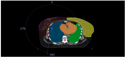
Figure 1: Represents the contours of PTV, heart, both lungs and contralateral breast
Planning methods and dose restrictions
Each treatment strategy uses Elekta linear accelerators (Infinity, Elekta) running at 6 MV photons to deliver 40.5 Gy in 15 fractions to the PTV. All treatment strategies attempted to minimize the exposure to OARs while covering at least 95% of the PTV by 95% of the recommended dose. Figure 2 shows VMAT strategies.
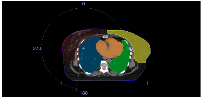
Figure 2: Represented the VMAT plan technique for lumpectomy
Evaluation tools
Dose volume histograms will be used to objectively analyse the PTV, heart, thyroid, and other critical normal tissues (lungs and the contralateral breast) as part of the treatment regimens, as shown in Figure 3.
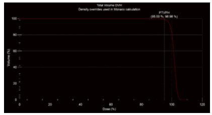
Figure 3: Dose distribution and DVH
Statistical analysis
For statistical analysis, IBM SPSS statistics (version 26) was used. In order to compare the mastectomy and lumpectomy plans, unpaired samples testing was performed. The threshold for statistical significance was established at 5% (P=0.05).
Results
According to Table 1 and Figure 4, the VMAT technique achieved optimal dose distribution in both lumpectomy and mastectomy conditions. P-value (0.151893287) however, for Axillary and Supraclavicular lymph node target (PTV AXI + SC), the results were almost the same PValue (0.603909676).
Tab. 1. The tumor's coverage (PTV (C.W) and PTV (AXI + SC))
| Tumor | Lumpectomy | Mastectomy | p-value |
|---|---|---|---|
| PTV AXI = SC | 99.69 ± 0.55 | 99.78 ± 0.17 | 0.603909676 |
| PTV C. W | 98.95 ± 0.46 | 98.44 ± 0.96 | 0.151893287 |
Figure 4: Represents coverage of the tumor PTV (C.W) & PTV (AXI+SC)
Table 2 and Figure 5 show that the dose delivered to the right lung in the case of lumpectomy is lower compared to the case of mastectomy were of P-value (0.000306278). Likewise, for the heart, Table 2 shows that the dose delivered in the case of Lumpectomy is lower compared to mastectomy, but did not significant and were of P-value (0.074810746). For the left lung, the administered dose
Tab. 2. Dose for Organ at Risk (OAR)
| Organs at Risks | Lumpectomy | Mastectomy | p-value |
|---|---|---|---|
| Thyroid | 18.14 ± 7.90 | 19.11 ± 4.62 | 0.732696042 |
| Contralateral Breast | 4.27 ± 0.90 | 3.85 ± 1.11 | 0.245976221 |
| Left Lung | 3.32 ± 0.50 | 4.16 ± 0.47 | 0.000246627* |
| Right Lung V20% | 24.79 ± 1.97 | 27.85 ± 1.63 | 0.000306278* |
| Right Lung Mean (Gy) | 12.87 ± 0.79 | 14.17 ± 0.79 | <0.00001 |
| Heart | 3.99 ± 0.66 | 4.52 ± 0.76 | 0.074810746 |
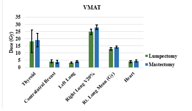
Figure 5: Represents dose for Organ at Risk (OAR)
in the case of lumpectomy is lower compared to that in the case of mastectomy P-value (0.000246627). Finally, the dose delivered to the contralateral breast was lower in the case of mastectomy compared to the case of lumpectomy P-value (0.245976221) but did not significant.
Discussion
The liver tissue is not homogeneous; it shows variability with characteristic geometry. This research affords a statistical analysis of apparently normal liver tissue of adult human in cases of adenocarcinoma. It was documented in the results of this study that recognizing the histological assessment of the apparently liver tissue could be evaluated quantitatively. The current study provides a database on which to make quantitative and qualitative statements about this tissue. The documentation of the normal criteria of this tissue could contribute in upcoming studies of the liver response to injury. Also, the results offer quantitative provision for a pathologist’s report to the physician accomplishment of the biopsy.
The literatures published did not discuss any of the data regarding morphometric parameters of the stromal-parenchymal components of the apparently normal liver tissue around adenocarcinomatous masses. The resulting morphometric data stromal-parenchymatous component reported by this study can be used to be compared with a control group in a study that may have a facility for obtaining normal liver biopsy may be from autopsy. The biopsy of liver is regarded as the best method for assessing the normality of liver tissue, however, this procedure is invasive. It is difficult to obtain normal liver biopsy from a volunteer. If such a comparison is performed, the answer to the question could research work on the apparently normal liver tissue surrounding pathology in the liver been considered normal, at least from histological point of view. Accordingly, histochemical techniques for staining of this apparently normal liver tissue may be considered as a valid tool to investigate predisposing factors leading to a specific pathology
Discussion
The goal of this study is to maintain and improve PTV dosage coverage while reducing the dose to the nearby normal tissues. One recent innovation in radiation therapy is VolumetricModulated Arc Therapy (VMAT), which protects healthy tissue while increasing coverage of the target volume. Although postoperative radiation for breast cancer has been shown to be effective, there are a number of potential side effects that could negatively impact patient quality of life and even their chance of survival. Significant long-term radiation side effects include second cancers, lymphoedema, brachial plexopathy, cardiac and lung damage, and lung and heart disease. Modern technology has reduced the risk of severe problems, which was formerly very significant with outdated, ineffective irradiation treatments. This study reviews current research [6]. This study demonstrated that in both situations (lumpectomy and mastectomy), clinical PTV was adequately covered by a dose, with a little advantage in the lumpectomy P-value (0.151893287) condition. Numerous papers have examined the possibility of pulmonary damage following radiotherapy for breast cancer, with incidence rates varying greatly and falling between 0% and 25%. The amount of lung that was exposed to radiation determines both immediate and long-term damage, Kahán Z and your colleagues reported that the risk of early and late irradiated lung sequelae is highly correlated with the patient's age, the size of the irradiated lung, and its dose. The grade of fibrosis increased with increasing lung volume treated to a radiation of 25 Gy. The current study indicated a considerable benefit in protecting the ipsilateral lung in the event of Lumpectomy P-value (0.000306278*), as shown in Table 2. This is in line with past a study that shown clinically symptomatic pneumonitis was uncommon when the V20 Gy of the ipsilateral lung was kept below 30% [7, 8].
Regarding the heart, since coronary artery disease is the most prevalent type of heart disease, it is likely that radiation operates, at least in part, by inducing or encouraging coronary atherosclerosis. However, the exact mechanism by which radiation causes heart disease is still unknown [9]. For the purpose of helping oncologists design the specific course of therapy for each woman, estimates of the absolute risks of radiation-related ischemic heart disease are required. The benefits of radiation in their entirety can then be weighed against these hazards [10].
The incidence of coronary events among breast cancer patients who received radiation between 1958 and 2001 is described by Darby et al. The truth of the matter is that radiation delivery methods have significantly advanced over time [11].
However, treatment plans in case of lumpectomy were better in protecting the heart compares with mastectomy but without any significant as shown in Table 2. This is consistent with earlier research that found that radiation exposure to the heart during breast cancer radiotherapy increased the risk of developing ischemic heart disease later on. Beginning a few years after exposure, and continuing for at least 20 years, this boost is proportionate to the average heart dose. Women with pre-existing cardiac risk factors are significantly more likely to experience side effects from radiation than other women [12].
Regarding the opposite breast, radiotherapy for breast cancer doesn't significantly reduce the likelihood of acquiring a second cancer there, which is already high [13]. Young patients with BC irradiated with breast tangential experience increased risk of CBC, especially in those with a positive family history of BC [14].Various studies evaluated the connection between radiation exposure and the risk of eventual primary cancer in the Contralateral Breast (CB) [15, 16].
Research conducted by Basco on more than 14,000 women undergoing breast cancer treatment in British Columbia found that the number of CB cancers was low (n=194, with only 30 occurring in women aged 50 to 64) because the IMRT technique reduced the dose delivered to the contralateral breast [17-20].
Conclusion
When comparing between (mastectomy and lumpectomy) VMAT has more successful results in case of dose reduction to the both lungs, while for PTV coverage this study showed that clinical PTV was covered by an adequate dose in both cases (lumpectomy, mastectomy) with a little advantage in the case of lumpectomy.
References
- Alwakeel AF, Al Musawi MS, Alabedi HH, Mohammed HJ. RETRACTED ARTICLE: Dosimetric assessment of IMRT treatment planning for unilateral breast cancer patient using Octavius phantom detector. Appl. Nanosci., 2023; 13:549
[Google Scholar][Cross Ref]
- Basco VE, Coldman AJ, Elwood JM, Young ME. Radiation dose and second breast cancer. Br J Cancer. 1985; 52:319-325.
[Google Scholar][Cross Ref]
- Baskar R, Lee KA, Yeo R, Yeoh KW. Cancer and radiation therapy: current advances and future directions. Int. J. Med. Sci., 2012; 9:193.
[Google Scholar][Cross Ref]
- Boice Jr JD, Harvey EB, Blettner M, Stovall M, Flannery JT. Cancer in the contralateral breast after radiotherapy for breast cancer. N Engl J Med. 1992; 326:781-785.
[Google Scholar][Cross Ref]
- Darby SC, Ewertz M, McGale P, Bennet AM, Blom-Goldman U et al. Risk of ischemic heart disease in women after radiotherapy for breast cancer. N Engl J Med. 2013; 368:987-998.
[Google Scholar][Cross Ref]
- De Benedetto D, Abdulcadir D, Giannotti E. Radiological anatomy of the breast. Ital J Anat Embryol. 2016:20-36.
[Google Scholar][Cross Ref]
- Boice Jr JD, Harvey EB, Blettner M, Stovall M, Flannery JT. Cancer in the contralateral breast after radiotherapy for breast cancer. N Engl J Med. 1992; 326:781-785.
[Google Scholar][Cross Ref]
- Ferlay J, Colombet M, Soerjomataram I. Estimating the global cancer incidence and mortality in 2018: GLOBOCAN sources and methods. Int J Cancer. 2019;144:1941-1953.
[Google Scholar][Cross Ref]
- Fisher B. Laboratory and clinical research in breast cancer—a personal adventure: the David A. Karnofsky memorial lecture. Cancer Res. 1980; 40:3863-3874.
[Google Scholar][Cross Ref]
- Fisher B, Anderson S, Bryant J. Twenty-year follow-up of a randomized trial comparing total mastectomy, lumpectomy, and lumpectomy plus irradiation for the treatment of invasive breast cancer. N Engl J Med. 2002; 347:1233-1241.
[Google Scholar][Cross Ref]
- Hooning MJ, Aleman BM, Hauptmann M. Roles of radiotherapy and chemotherapy in the development of contralateral breast cancer. J Clin Oncol. 2008;26:5561-5568.
[Google Scholar][Cross Ref]
- Krengli M, Sacco M, Loi G. Pulmonary changes after radiotherapy for conservative treatment of breast cancer: a prospective study. Int J Radiat Oncol* Biol* Phys. 2008; 70:1460-1467.
[Google Scholar][Cross Ref]
- Low C, Chen Y, Wu M. Understanding the determinants of cloud computing adoption. Ind Manag Data Syst. 2011; 111:1006-1023.
[Google Scholar][Cross Ref]
- Mestres A, Rodriguez-Natal A, Carner J. Knowledge-defined networking. ACM SIGCOMM Comput Commun Rev. 2017; 47:2-10.
[Google Scholar][Cross Ref]
- Senkus-Konefka E, Jassem J. Complications of breast-cancer radiotherapy. Clin Oncol. 2006; 18:229-235.
[Google Scholar][Cross Ref]
- Shimm DS. Perez and Brady's Principles and Practice of Radiation Oncology. Int J Radiat Oncol Biol Phys. 2008;72:1268.
[Google Scholar][Cross Ref]
- Sun YS, Zhao Z, Yang ZN. Risk factors and preventions of breast cancer. Int J Biol Sci. 2017; 13:1387.
- Taylor CW, Kirby AM. Cardiac side-effects from breast cancer radiotherapy. Clin Oncol. 2015; 27:621-629.
[Google Scholar][Cross Ref]
- Taylor CW, McGale P, Darby SC. Cardiac risks of breast-cancer radiotherapy: a contemporary view. Clin Oncol. 2006; 18:236-246.
[Google Scholar][Cross Ref]
- Wang R, Shen J, Yan H. Dosimetric comparison between intensity-modulated radiotherapy and volumetric-modulated arc therapy in patients of left-sided breast cancer treated with modified radical mastectomy: CONSORT. Medicine. 2022; 101.


