Research Article - Onkologia i Radioterapia ( 2020) Volume 14, Issue 4
Analysis of dosimetric parameter on craniospinal irradiation with Helical Tomotherapy (HT), 3D Conformal Radiotherapy (3DCRT), and Intensity Modulated Radiotherapy (IMRT)
Fauzan Herdian, Anak Agung Sagung Ari Lestari, Vito Filbert Jayalie, Handoko, Wahyu Edi Wibowo, Muhammad Djakaria and Soehartati Gondhowiardjo*Soehartati Gondhowiardjo, Department of Radiation Oncology, Faculty of Medicine University of Indonesia, Dr. Cipto Mangunkusumo Hospital,Jakarta, Indonesia, Tel: 0878-8857-6057, Email: gondhow@gmail.com
Received: 14-Jun-2020 Accepted: 25-Jun-2020 Published: 03-Jul-2020
Abstract
Traditional craniospinal irradiation consists of a large treatment area divided into several fields that can create dose overlaps in the inter-field junction. This analysis compares the dosimetric parameter of craniospinal irradiation with HT, 3DCRT, and LINAC-based IMRT and find the optimal technique in terms of dose distribution, organ-sparing, and body radiation exposure.
In our hospital, 3DCRT, IMRT, and HT plan were made from CT data of 10 patients indicated for craniospinal irradiation, with a total dose of 36 Gy in 20 fractions. Cranial and spinal PTV coverage was evaluated using the Conformity Index (CI) and Homogeneity Index (HI). Dose received by critical organs, body-wide radiation exposure, number of Monitor Units (MU), and beam on duration were recorded and compared. In cranial PTV, HT and IMRT had better HI and CI compared to 3DCRT with no significant difference between IMRT and HT. In spinal PTV, HT had better HI and CI compared to IMRT and 3DCRT. 3DCRT has the highest mean dose in most of the critical organs, while HT has the highest whole-body radiation exposure, highest number of MU, and the longest beam on duration. For doses in inter-field junction, there is no statistically significant difference between 3DCRT and IMRT techniques. HT technique achieved the highest HI and CI but also had the highest body-wide radiation exposure, highest MU number, and longest beam on duration, in contrast to 3DCRT Proper consideration of the technique used in craniospinal irradiation is important to prevent late side effects, such as secondary malignancy.
Keywords
Dosimetry, craniospinal, 3DCRT, IMRT, helical tomotherapy
Introduction
Craniospinal irradiation is a therapeutic option for patients with central nervous system malignancy who are at risk of developing Cerebrospinal Fluid (CSF) dissemination. The goal of craniospinal irradiation is to give doses as homogenous as possible throughout the subarachnoid space, covering the entire cranial cavity, spinal canal, to the tip of the thecal sac [1-3]. Due to the wide irradiation area, dose to critical organs must be taken into account in craniospinal irradiation. Also, most patients in need of craniospinal irradiation are children so the risk of secondary malignancy must also be considered [4].
Previously, craniospinal irradiation was performed using conformal Two Dimensional (2D) and Three Dimensional- Conformal Radiotherapy (3DCRT) techniques on LINAC machines. Currently, Intensity-Modulated Radiation Therapy (IMRT) technique can also be used due to better dose distribution on Planning Target Volume (PTV) and better sparing of critical organs. Yet, both 3DCRT and IMRT techniques still have obstacles in the field gaps. With the current technological developments, IMRT with Helical Tomotherapy (HT) is one of the emerging modalities for craniospinal irradiation, and it is expected to improve the dose distribution due to its ability to irradiate the treatment field in a single pass [4, 5]. A study conducted by Studenski et al. at East Carolina University showed that IMRT and VMAT produced more homogenous doses at target volumes and fewer doses received by critical organs compared to 3DCRT. Yet, similar to IMRT HT, the low doses area became wider [6].
Cipto Mangunkusumo General Hospital is the national referral hospital in Indonesia and currently the only hospital in Indonesia with HT machine. Therefore, we planned to conduct this analysis of craniospinal irradiation dosimetry with 3D-CRT, LINAC-based IMRT and tomotherapy-based IMRT techniques as a reference to guide us in determining the appropriate craniospinal irradiation technique for indicated patients [7, 8].
Materials and Methods
Patients
This study was conducted from January to February 2018 at Cipto Mangunkusumo Hospital Department of Radiotherapy. We used Computed Tomography (CT) data of 10 patients indicated for craniospinal irradiation based on our medical record from 2016 to 2018 [9-11].
Planning
3DCRT and IMRT plan consist of the cranial and spinal fields. In the 3DCRT plan, cranial fields were irradiated with 2 beams from the opposing lateral direction with collimation angle adjusted during the planning and the spinal field irradiated from the posterior (180° angle). In the IMRT plan, the cranial field was irradiated using 6 beam directions and the spinal field using 3 beam directions. Dose calculation for both techniques was done using Anisotropic Analytical Algorithm (AAA) with 2.5 mm spatial resolution. HT plan used a 5 cm field width, 0.43 normal grid pitch with 2.0 modulation factor [12, 13]. Quality assurance (QA) was performed on the plan by an experienced medical physicist. Dosimetric parameters of the Dose Volume Histogram (DVH) print out were recorded in tables. Analysis of variables was carried out using the IBM SPSS statistics version 21.0. Characteristics were displayed descriptively. The distribution of HI, CI, D98%, D2%, D50%, dose in critical organs and whole-body radiation exposure, beam duration and number of MU in each external irradiation technique were analysed using statistical tests.
Results
10 patients (8 paediatric and 2 adult patients) were involved. Mean age was 15.80 years with the lowest age was 5 years and the highest was 49 years as shown in (Table 1).
| No | Age (years) | Sex |
|---|---|---|
| 1 | 12 | Male |
| 2 | 6 | Male |
| 3 | 49 | Female |
| 4 | 11 | Male |
| 5 | 16 | Male |
| 6 | 5 | Male |
| 7 | 7 | Female |
| 8 | 13 | Female |
| 9 | 12 | Female |
| 10 | 27 | Male |
Mean age: 15, 80
Standard deviation: 12, 7
Minimum-Maximum: 5-49
Table 1. Basic characteristics of patients
Cranial PTV
Mean cranial PTV dosimetry parameters between 3DCRT, IMRT and HT techniques in craniospinal irradiation can be seen in (Table 2).
| 3D CRT | IMRT | HT | p-value 3D CRT vs IMRT | p-value 3D CRT vs HT | p-value IMRT vs HT | ||||
|---|---|---|---|---|---|---|---|---|---|
| Parameter | Mean | SD | Mean | SD | Mean | SD | |||
| D98% | 34.75 | 1.31 | 34.58 | 0.62 | 33.77 | 0.68 | 0.729 | 0.044 | 0.011 |
| D50% | 37.4 | 0.17 | 36.27 | 0.23 | 35.97 | 0.04 | <0.001 | <0.001 | 0.003 |
| D2% | 39.5 | 0.63 | 37.31 | 0.53 | 37.15 | 0.26 | 0.005 | 0.005 | 0.508 |
| HI | 0.13 | 0.04 | 0.08 | 0.03 | 0.09 | 0.02 | 0.017 | 0.037 | 0.005 |
| CI | 1.25 | 0.12 | 1.09 | 0.05 | 1.09 | 0.06 | <0.001 | 0.008 | 0.882 |
Table 2: Mean cranial PTV dosimetry parameters among 3D-CRT, IMRT SS and HT techniques in craniospinal irradiation
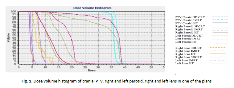
The mean HI was higher in the IMRT technique (0.08) compared to 3D-CRT technique (0.13). There were no significant differences in mean HI between IMRT and HT techniques. The mean CI value in IMRT and HT techniques was 1.09 and in 3DCRT technique was 1.25 (p<0.05). The difference of cranial PTV between each technique in Dose- Volume Histogram (DVH) can be seen in Figure 1.

Figure 1: Dose volume histogram of cranial PTV, right and left parotid, right and left lens in one of the plans
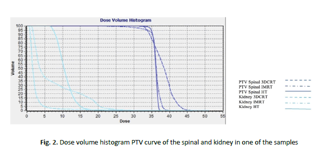
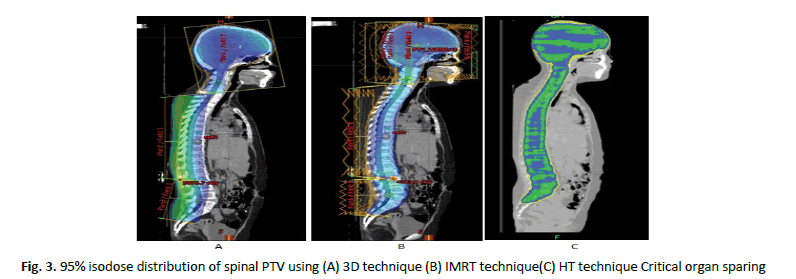
HT has the highest mean HI (0.05) compared to IMRT (0.17) and 3DCRT (0.32). HT also has the highest mean CI (1.09) compared to IMRT (1.20) and 3DCRT (2.26). There was a significant difference in mean HI and mean CI between the three techniques. The difference of spinal PTV can be seen in (Figure 2). 95% isodose distribution of spinal PTV between each technique can be seen in (Figure 3).

Figure 2: Dose volume histogram PTV curve of the spinal and kidney in one of the samples

Figure 3: 95% isodose distribution of spinal PTV using (A) 3D technique (B) IMRT technique(C) HT technique Critical organ sparing
Between each technique, HT was able to achieve the lowest critical organ mean dose in both lens (7.29 and 6.93 Gy), eyes (14.62 and 14.30 Gy), both parotids (15.04 and 14.60 Gy), thyroid (21.69 Gy), V25 of heart (0.55%), and medulla spinalis (36.95 Gy). 3DCRT technique, on the other hand, able to reduce mean dose in both submandibular glands (7.90 and 9.28 Gy), oral cavity (5.23 Gy), V20 of the left lung (3.04%), and both kidneys (3.60 Gy), while IMRT produce the lowest critical organ mean dose in V20 of right lung (7.18%), ovarium (1.04 Gy in 4 female patients), and testes (0.22 Gy in 6 male patients) (Table 3).
| 3D CRT | IMRT | HT | p-value 3D CRT vs IMRT | p-value 3D CRT vs HT | p-value IMRT vs HT | ||||
|---|---|---|---|---|---|---|---|---|---|
| Parameter | Mean | SD | Mean | SD | Mean | SD | |||
| D98% | 31.51 | 1.24 | 32.16 | 1.02 | 35.06 | 0.47 | 0.27 | <0.001 | <0.001 |
| D50%* | 37.4 | 0.52 | 36.57 | 0.22 | 36.1 | 0.12 | 0.005 | 0.005 | 0.005 |
| D2%* | 43.34 | 1 | 38.32 | 0.46 | 36.84 | 0.23 | 0.005 | 0.005 | 0.005 |
| HI | 0.32 | 0.06 | 0.17 | 0.03 | 0.05 | 0.02 | <0.001 | <0.001 | <0.001 |
| CI | 2.26 | 0.39 | 1.2 | 0.07 | 1.09 | 0.07 | <0.001 | <0.001 | 0.004 |
*Indicates an abnormal data distribution and is analyzed by Wilcoxon's non-parametric test
Table 3. Mean spinal PTV dosimetry parameters among 3D-CRT, IMRT SS and HT techniques in craniospinal irradiation
Whole-body radiation exposure
Whole-body radiation exposure is defined as a dose obtained by body area outside the PTV. We measured the theme and dose of the body and body volume receiving 5 Gy (Table 4).
| 3D CRT | IMRT | HT | p-value 3D CRT vs IMRT | p-value 3D CRT vs HT | p-value IMRT vs HT | |||||
|---|---|---|---|---|---|---|---|---|---|---|
| Parameter | Mean | SD | Mean | SD | Mean | SD | ||||
| Dmax of Lens (Gy) | Right | 7.29 | 1.66 | 9.38 | 0.83 | 7.29 | 1.13 | <0.001 | 0.991 | 0.001 |
| Left | 5.95 | 1.45 | 9.36 | 0.74 | 6.93 | 1.11 | <0.001 | 0.066 | <0.001 | |
| Dmean of Eyes (Gy) | Right | 17.83 | 3.42 | 17.53 | 1.97 | 14.62 | 1.57 | 0.753 | 0.008 | 0.002 |
| Left | 16.96 | 2.8 | 17.76 | 2.55 | 14.3 | 1.29 | 0.521 | 0.031 | 0.002 | |
| Dmean of Parotid glands (Gy) | Right | 30.83 | 6.03 | 17.55 | 1.17 | 15.04 | 2.31 | 0.005 | 0.005 | 0.022 |
| Left | 31.47 | 5.22 | 17.35 | 1.3 | 14.6 | 2.27 | 0.005 | 0.005 | 0.009 | |
| Dmean of Submandibular glands (Gy) | Right | 7.9 | 4.1 | 15.7 | 1.85 | 17.87 | 1.19 | <0.001 | <0.001 | 0.003 |
| Left | 9.28 | 5.8 | 17.12 | 2.17 | 18.34 | 2.52 | 0.007 | 0.007 | 0.203 | |
| Dmeanof oral cavity (Gy) | 5.23 | 2.48 | 11.83 | 2.37 | 18.33 | 1.48 | 0.005 | 0.005 | 0.005 | |
| Dmean of thyroid (Gy) | 27.43 | 3.68 | 22.87 | 1.93 | 21.69 | 3.33 | 0.007 | 0.009 | 0.646 | |
| V25 of Heart (%) | 29.95 | 15.11 | 0.69 | 1.21 | 0.55 | 0.67 | 0.005 | 0.005 | 0.767 | |
| V20 of Lung (%) | Right | 9.96 | 4.81 | 7.18 | 2.61 | 11.59 | 2.04 | 0.038 | 0.251 | <0.001 |
| Left | 3.04 | 2.34 | 3.82 | 1.97 | 8.32 | 1.97 | 0.17 | <0.001 | <0.001 | |
| Dmean of Kidney (Gy) | 3.6 | 2.05 | 8.23 | 2.24 | 13.23 | 1.92 | <0.001 | <0.001 | <0.001 | |
| Dmean of Testes (Gy) | 0.28 | 0.29 | 0.22 | 0.15 | 0.27 | 0.09 | 0.465 | 0.911 | 0.267 | |
| Dmean of Ovarium(Gy) | 1.3 | 0.61 | 1.04 | 0.96 | 2.61 | 1.72 | 0.184 | 0.7 | 0.315 | |
| Dmax of Spinal cord | 43.49 | 1.87 | 43.08 | 2.79 | 36.95 | 0.14 | 0.646 | <0.001 | <0.001 | |
Table 4. Mean critical organdosimetry parameters among DCRT, IMRT SS, and HT techniques in craniospinal irradiation
Table 5 describes dosimetric parameters of whole-body radiation exposure between 3DCRT, IMRT SS and HT techniques in Craniospinal Irradiation, which shows that the highest D mean was found in HT technique (9.62+1.11) and the lowest in 3DCRT technique (6.27+1.53). A statistically significant difference of D mean was obtained from the whole-body radiation exposure among the three techniques. Whole-body volume which received 5 Gy dose was found the highest in HT technique (71.96+6.86) and the lowest in 3DCRT technique (22.40+4.10).
| 3D CRT | IMRT | HT | p-value 3D CRT vs IMRT | p-value 3D CRT vs HT | p-value IMRT vs HT | ||||
|---|---|---|---|---|---|---|---|---|---|
| Parameter | Mean | SD | Mean | SD | Mean | SD | |||
| Dmean (Gy) | 6.27 | 1.53 | 7.39 | 1.41 | 9.62 | 1.11 | 0.022 | <0.001 | 0.001 |
| V5 (percentage) | 22.4 | 4.1 | 43.86 | 6.05 | 71.96 | 6.86 | <0.001 | <0.001 | <0.001 |
Table 5. Mean whole body radiation exposure dosimetry parameters among 3D CRT, IMRT SS and HT techniques in craniospinal irradiation
Number of MU and beam on time
Monitor Unit (MU) is the amount of charge recorded in the ionization chamber on the head of a linear accelerator which correlates with a dose of 1 cGy delivered to a water phantom under reference conditions. The number of MU, combined with the beam on time, represents the length of treatment time and radiation exposure to the body per session. Table 6 shows the mean number of Monitor Unit (MU) parameters between 3D-CRT, IMRT SS and HT techniques in craniospinal irradiation. Our results show that the lowest number of MU was found in the 3DCRT technique (524.60+120.99) and the highest in the HT technique (6514.00+1407.22). The shortest beam on time was also found in the3D-CRT technique (1.36+0.37) and the longest in the HT technique (7.77+1.65).
| 3D CRT | IMRT | HT | p-value 3D CRT vs IMRT | p-value 3D CRT vs HT | p-value IMRT vs HT | ||||
|---|---|---|---|---|---|---|---|---|---|
| Parameter | Mean | SD | Mean | SD | Mean | SD | |||
| MU* | 524.6 | 120.99 | 1518.4 | 229.71 | 6514 | 1407.22 | 0.005 | 0.005 | 0.005 |
| Beam Duration* | 1.36 | 0.37 | 3.79 | 0.59 | 7.77 | 1.65 | 0.005 | 0.005 | 0.005 |
* Indicates an abnormal data distribution that the author analysed with Wilcoxon's non-parametric test
Table 6. Mean number of monitor unit and beam on time parameters among 3D CRT, IMRT SS and HT techniques in craniospinal irradiation
There was a statistically significant difference in the number of MU and the duration of beam-on time among the three techniques.
Dose on junction
In the 3D-CRT technique, the mean Dmax at the upper (cranial-spinal) and lower (spinal-spinal) junctions were higher than those in the IMRT technique. However, as seen in the statistical analysis of Table 7, there were no statistically significant differences between the two techniques. HT technique did not create any junction since the craniospinal area was irradiated in a single field.
| 3D CRT | IMRT | p-value 3D CRT vs IMRT |
|||
|---|---|---|---|---|---|
| Parameter | Mean | SD | Mean | SD | |
| Dmax of upper junction | 44.46 | 3.05 | 42.67 | 2.39 | 0.52 |
| Dmax of lower junction | 47.01 | 1.33 | 45.53 | 1.36 | 0.05 |
| Dmin of upper junction | 21.76 | 7.5 | 21.65 | 1.65 | 0.89 |
| Dmin of lower junction | 25.85 | 7.39 | 17.42 | 3.87 | 0.03 |
Table 7. Dose on Junction
Discussion
In this dosimetric study, we found that modern radiotherapy techniques (IMRT and HT) show better dose distribution for cranial PTV compared to 3DCRT. The IMRT and HT technique shows the lowest HI value (0.08 and 0.09, respectively) compared to 3DCRT (0.13). For CI value, IMRT and HT techniques also able to achieve better conformity than 3DCRT, confirm in gan earlier study that HI and CI achieved using 3D-CRT technique was less ideal compared to IMRT [14-16]. Unlike cranial PTV, Spinal PTV using HT technique was able to achieve the best dose conformity and homogeneity compared to other techniques (1.09 and 0.05, respectively). To apply this results in clinical practice, proper immobilization during the simulation and correct volume delineation are a prerequisite to gain the benefit of these highly conformal techniques.
In terms of critical organ sparing in the head and neck area, there are mixed results from each technique. HT techniques can spare the lens, eyes, parotid glands, and thyroid, but unable to spare submandibular glands and oral cavity, of which 3DCRT technique was able to spare. The difference in mean dose spared from the organ, especially in parotid glands, submandibular glands, oral cavity, and thyroid are also quite high (more than 5 Gy). The beam in the cranial region with 3DCRT technique came from opposing lateral direction, in which parotid gland block could not be performed with MLC because it would reduce the PTV dose, therefor increasing its received dose. This result is also confirmed in another study where radiation dose to salivary glands as a critical organ was higher in 3DCRT compared to HT [17]. Several works also had confirmed our results in thyroid sparing, with IMRT and HT able to achieve lower mean dose compared to 3DCRT [17, 18]. For oral cavity and submandibular glands, the 3DCRT technique can achieve smaller mean dose due to lower cranial-spinal junction set by the author (5th-6th cervical vertebrae), enabling both organs to be blocked by the MLC [8].
Heart and lungs are important organs which function could deteriorate due to the late effect caused by radiation therapy. In our study, we found that IMRT and HT can significantly spare the heart, represented in mean heart volume receiving 25 Gy, compared to 3DCRT technique. On the other hand, lung volume which received 20 Gy was higher in the HT technique compared to 3DCRT and IMRT techniques. Although both results are still within our constraint, whether that received doses could cause significant acute or late toxicity are need to be analysed in a different study.
For the abdominal area, we found that both kidneys received a significant mean dose with the HT technique compared to the other techniques. Another study has also shown that in craniospinal irradiation with 3DCRT and HT techniques, the dose obtained by both kidneys are higher in HT technique [17]. For reproductive organs, the mean dose obtained by testes and ovaries were not significantly different from one technique to another, although the dose on the ovary seemed higher in the use of HT technique (2.61 Gy) compared to 3DCRT (1.30 Gy) and IMRT (1.04 Gy). However, since the function of those organs could be affected even by small doses of radiation, we need to be prudent in choosing the right techniques, especially in paediatric patients where the preservations of their reproductive capabilities are an important goal.
Although the spinal cord is our target in craniospinal irradiation, we found that HT can limit the maximum dose and give the closest to our prescribed ones (36.95 ± 0.14 Gy). The dose range of HT also close to zero, which means these results will be reproducible between various patients.
Whole-body radiation exposure is another important factor that should be considered in selecting a radiotherapy technique, especially in patients with good prognosis after treatment, because of the late effects that a rise could greatly affect their quality of life. In this study, we found that the mean dose for whole-body exposure was the highest in the HT technique compared to 3DCRTand IMRT techniques. Likewise, the volume of the body receiving 5 Gy, in HT, IMRT and 3D were 71.96% ± 6.86%, 43.86% ± 6.05%, and 22.40% ± 4.10%, respectively. This is due to HT multiple, low dose beam which creates a “dose bath” to the area surrounding the PTV, and this is also shown in a similar study where the body volume receiving doses of 4 Gy in HT techniques is around 50%-57%, while 3DCRT techniques are 25%-27% [13].
The 3DCRT technique was the most efficient in MU number and beam on time compared to IMRT and HT techniques, with MU number of 524.60 ± 120.99 and beam on time of 1.36 minutes, due to less complicated plan. Whereas HT had the highest MU number of 6514.00 ± 1407.22 and the longest beam on time of 7.77 ± 1.65 minutes. Several studies had stated that the conventional 3DCRT technique was the most efficient in saving MU and provided a shorter beam on time compared to advanced techniques such as IMRT and VMAT [15, 18]. The advantage of less MU and beam on time is shorter treatment time per session and less secondary scatter from treatment machine, although this should not prevent us from choosing more advanced techniques to plan and irradiate patients with extensive tumor extension, where those highly conformal techniques could bring more benefit.
Dose at the junction must be carefully planned to prevent hotspot or coldspot. Any hotspots that occur means there is a risk of side effects to the tissue outside PTV, while cold spots inside PTV mean recurrence risk inside the treatment field. In this study, the upper junction Dmax value using 3DCRT technique was higher than IMRT technique, although there were no statistically significant differences between the Dmax values of the upper and lower junction using both techniques. The upper junction Dmin value for the 3DCRT technique was also not significantly different from IMRT, while the lower junction Dmin value in 3DCRT technique was significantly higher from the IMRT technique. The advantage of the HT technique is that it does not require the production of junctions so it creates more conformal dose distribution with no risk of hotspots or cold spots in their radiated area.
Conclusion
In conclusion, HT techniques gives a superior dose distributions compared to IMRT and 3DCRT at the cost of higher integral dose to tissues outside the PTV. This, combined with the higher MU and longer beam on time, means a potentially higher risk of secondary malignancy in specific patients such as patients at a very young age and with a genetic predisposition to certain cancer. Proper consideration in choosing radiation techniques for craniospinal irradiation between different patients is needed. Further research into novel craniospinal irradiation techniques, such as using proton therapy, is also important to limit the acute and late treatment toxicities.
Conflict of Interest
He authors declare that they have no conflict of interest in this study.
Statement of Human Rights
All procedures performed in studies involving human participants were in accordance with the ethical standards of the Institutional Review Board (IRB) and with the 1964 Helsinki declaration and its later amendments or comparable ethical standards.
Informed Consent
Informed consent was obtained from all individual participants included in the study.
Acknowledgment
None
References
- Freeman CR, Farmer JP, Taylor RE. Central nervous system tumour in children: in perez and bradys principles and practices of radiation oncology. Philadelphia: Lippincot Williams and Wilkins. 2013:1632-1655.
- Cancer C, Incidence AC. Pediatric radiotherapy planning and delivery overview of this presentation childhood cancers are different than adult cancers relative number of cancers by RT vs . Chemo vs . Both Hodgkin’s. Disease Induction of Second Cancer for Pediatric RT Treatment. 2013;1-14.
- Sugie C, Shibamoto Y, Ayakawa S, Mimura M, Komai K, et al. Craniospinal irradiation using helical tomotherapy: evaluation of acute toxicity and dose distribution. Technol Cancer Res Treat. 2011;10:187-195.
- Oliveria F, Lima F, Vilela E. Cranial-spinal junction and surrounding organs in medulloblastoma therapy : a dosimetric assessment between different techniques. Nucl Energy. 2018;1-6.
- Sharma SD, Gupta T, Jalali R, Master Z, Phurailatpam RD, et al. High-precision radiotherapy for craniospinal irradiation: evaluation of three-dimensional conformal radiotherapy, intensity-modulated radiation therapy and helical tomotherapy. Br J Radiol. 2009;82:1000-1009.
- Studenski M, Harrison A, Xiao Y, BiswasT. IMRT for craniospinal irradiation : a dosimetric comparison. Bodine J. 2010;3:1-17.
- Olch AJ. Pediatric radiotherapy, planning and treatment. J Chem Info Modeling. 2013;53:1-374.
- Michalski JM, Klein EE, Gerber R. Method to plan, administer, and verify supine craniospinal irradiation. J Appl Clin Med Phys. 2002;3:310-316.
- Combs SE, Lu JJ, Lee NY, Lu JJ. Target volume delineation for conformal and intensity-modulated radiation therapy. Radiation Oncol. 2015.
- Dobbs J, Barret A, Morris S, Roques T. Practical radiotherapy planning. 2nd ed. London: British Library Cataloguing in Publication Data. 2013.
- Kumar G, Yadav V, Singh P, Bhowmik KT. Radiation-Induced malignancies making radiotherapy a “ two-edged sword ”: a review of literature. World J Oncol. 2017;8:1-6.
- Baisden J, Benedict S,Sheng K,Read P,Larner J. Helical tomotherapy in the treatment of central nervous system metastasis. Neurosurg Focus. 2007;22:E8.
- Kunos CA, Dobbins DC, Kulasekere R, Latimer B, Kinsella T. Comparison of helical tomotherapy versus conventional radiation to deliver craniospinal radiation. Technol Cancer Res Treat. 2008;7:227-233.
- Supawongwattana B, Hoonghual T, Chitapanarux I, Wanwilairat S, Traisathit P. Dosimetric comparison of Helical Tomotherapy (HT) with intensity modulated radiotherapy (IMRT), Three- Dimension Conformal Radiotherapy (3D-CRT) and conventional two-Dimension Radiotherapy (2D) for Craniospinal Axis Irradiation (CSI). Chiang Mai Med J. 2015;54:17-28.
- Myers P, Stathakis S, Gutiérrez AN, Esquivel C, Mavroidis P, et al. Dosimetric comparison of Craniospinal Axis Irradiation (CSI) treatments using helical tomotherapy, smartarctm, and 3d conventional radiation therapy. Clin Eng Radiat Oncol. 2013;2:30-38.
- Srivastava R, Saini G, Sharma PK, Chomal M, Aagarwal A, et al. A technique to reduce low dose region for Craniospinal Irradiation (CSI) with rapidarc and its dosimetric comparison with 3D Conformal Technique (3DCRT). J Cancer Res Ther. 2015;11:488-491.
- Sugie C, Shibamoto Y, Ayakawa S, Mimura M, Komai K, et al. Craniospinal irradiation using helical tomotherapy: evaluation of acute toxicity and dose distribution. Technol Cancer Res Treat. 2011;10:187-195.
- Studenski MT, Shen X, Yu Y, Xiao Y, Shi W, et al. Intensity-modulated radiation therapy and volumetric-modulated arc therapy for adult craniospinal irradiation-a comparison with traditional techniques. Med Dosim. 2013;38:48-54.


