Case Report - Onkologia i Radioterapia ( 2023) Volume 17, Issue 8
Acute renal hemorrhage as a result of spontaneous rupture of angiomyolipoma
Arber Neziri1,2, Xhevdet Cuni2, Hasan Gashi1,2*, Liridon Selmani2 and Ilir Miftar22Urology Clinic, University Clinical Centre of Kosovo, Pristina, Kosovo
Hasan Gashi, Alma Mater European Campus College "Rezonanca", and Urology Clinic, University Clinical Centre of Kosovo, Pristina, Kosovo, Email: hasan.gashi@rezonanca-rks.com
Received: 26-May-2023, Manuscript No. OAR-23-100102; Accepted: 15-Sep-2023, Pre QC No. OAR-23-100102 (PQ); Editor assigned: 03-Jun-2023, Pre QC No. OAR-23-100102 (PQ); Reviewed: 18-Jul-2023, QC No. OAR-23-100102 (Q); Revised: 25-Aug-2023, Manuscript No. OAR-23-100102 (R); Published: 03-Sep-2023
Abstract
Introduction: Angiomyolipomas are benign tumours that are made up of a mixture of fatty tissue, thick blood vessels and smooth muscle in different proportions. Angiomyolipomas are found as solitary and multiple tumours of different sizes, they can be over 20 cm in diameter Symptomatic angiomyolipomas over 4 cm are potentially dangerous due to spontaneous rupture, nephrectomy is indicated in case of heavy bleeding which cannot be stopped or when the tumour involves the kidney.
Objective: Presentation of a rare case of spontaneous rupture of renal angiomyolipoma and subsequent development of haemorrhagic shock as well as adequate treatment.
Case Presentation: Patient K. K. born in 1974, initially with: 23.01.2017 at 02.30 min. admitted to abdominal surgery due to severe colic abdominal, which started in the periumbilical region and then spread throughout the abdomen. Due to the deterioration of the condition, it is decided to intervene surgically, since the urography suspected a ruptured angiomyolipoma, the patient was urgently admitted to the operating room After the application of nephrectomy, the bleeding stops and the patient's condition begins to stabilize.
Conclusions: Although benign tumours, angiomyolipomas can be quite dangerous tumours, especially angiomyolipomas over 4 cm, which rupture not infrequently.
Keywords
UCCK -prishtina, renal trauma, renal rupture, nephrectomia , renoraphia
Introduction
Angiomiolipomas are benign tumours that are constructed by mixing fatty tissue, thick blood vessels and smooth muscles in different proportions [1].
Participate in hamartoma, which consists of abnormal mixing tissue that is normally in the given organ. The muscular tissue has an embryonic appearance [1]. Symptomatic angiomyolipoma above 4 cm are potentially dangerous due to spontaneous rupture, nephrectomy is indicated in case of severe bleeding which cannot be stopped otherwise or when the tumour includes the entire kidney [2].
Almost one-third of the patients are reported with abdominal symptoms acutely due to kidney haemorrhage and peritoneal tissue [3-5]. Angiomiolipomas are solitary and multiple tumours with different sizes, in some cases can be over 20 cm in diameter [6]. Angiomiolipomas also appear as isolated kidney tumours. These sporadic cases are most common in females in the 5th and 6th decades, appearing in the form of unilateral tumours located in the kidney pole [7-9].
Angiomiolipomas are found in 50%-80% of patients with tuberous sclerosis, in these cases they are; Multiple, bilateral and often asymptomatic [9, 10]. Tuberous sclerosis is a hereditary disease that is characterized by; Epilepsy, mental retardation, sebaceous adenoma and hamartomas in various organs [10, 11].
Purpose of the study
Presentation of a rare case of spontaneous rupture of renal angiomyolipoma and consequent development of haemorrhagic shock and adequate treatment.
Case Presentation
Patient K. K. born in 1974, initially on: 23 .01.2017 at 02:30 AM was accepted in abdominal surgery due to strong abdominal colic,which started in the periumbilical region and then spread to the entire abdomen.
Biochemical analyzes were erythrocytes 3.72 , hematocrit 33 , hemoglobin 106 g / l, leucocytes-17.2 , urea-7.2 creatinine-72 , total bilirubin 1.2 m mol/l amylase / 49 mmol / l.
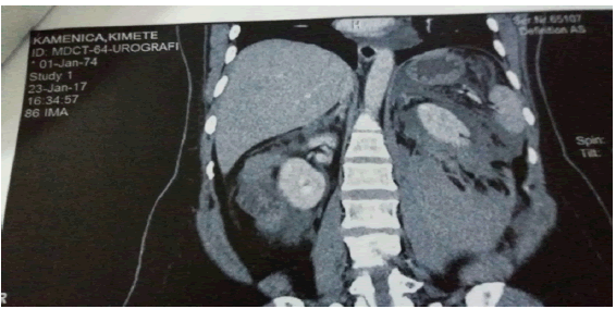
After the realization of abdominal CT and pelvic CT on January 23, 2017 at 16:34 and this image was obtained (Figure 1).

Figure 1: A CT scan showing the hematoma from the lower pole and the left renal side of the kidney
In the clinic the patient was treated with; Infusion, antibiotic, antiemetic, H2 blockader and analgesic, and 4 blood doses.
Despite receiving 4 doses of blood, haematocrit fell to 22 and Hgb to 60 g / l.
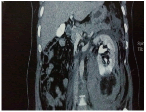
Due to the deterioration of the health condition I decided to intervene surgically because in the CT urography was suspected for being ruptured angiomyolipoma and on the patient was urgently placed in the theatre room in Figure 2.

Figure 2: A CT scan showing massive hematoma from the lower pole of the kidney
Results and Discussion
The patient intra-operative received 2 doses of plasma and 5 other doses of blood.
Operative approach is made, Op: Median laparotomy. Medial positioning of the descending colon that enables us to approach the hilus and take care of the hilus vessels. Once the tumour had a size of 13 by 10 cm+12 cm, the length of the kidney.
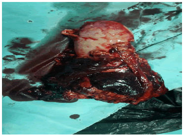
We also applied a transverse cut on the abdomen wall for easier access to the hilus. After application of nephrectomy the bleeding stops and the condition of the patient begins to stabilize (Figure 3).
Figure 3: A photo showing massive hematoma with kidney after nephrectomy.
Outcome
After surgery, the patient was extubated and sent to the Urological Clinic. Postoperatively patient was treated with infusions, transfusions, antibiotics and analgesics, and her condition was improved so that on the 5th postoperative day, the patient was released at home, in good condition and with improved laboratory analysis.
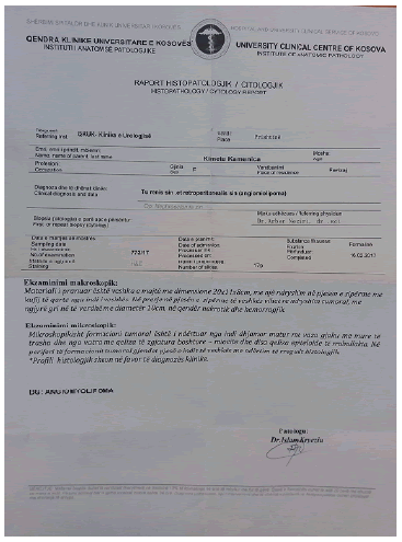
Histopathologic analysis of the tumour: angiomyolipoma is mentioning in the Figure 4.
Figure 4: A photo showing histopathologic diagnosis of tumour
Conclusion
Renal-Angio Myo Lipoma (AML) is a benign neoplasm of a hamartomata’s origin that presents lesions occasionally associated with tuberous sclerosis, or as a single unilateral lesion.
Usually are asymptomatic, but in some cases can manifest the following clinical triad: abdominal pain, palpable mass and haematuria. In our case massive retroperitoneal haemorrhage was found to be the cause, produced by spontaneous rupture of an angiomyolipoma in the left kidney. Despite being benign tumours, angiomiolipomas can be very dangerous tumours, especially angiomiolipomas over 4 cm, which can rupture very often.
References
- Eble JN. Angiomyolipoma of kidney. InSeminars Diagn Pathol. 1998; 15: 21-40.
- Neumann HP, Schwarzkopf G, Henske EP. Renal angiomyolipomas, cysts, and cancer in tuberous sclerosis complex. InSeminars Pediatr Neurol. 1998; 5: 269-275.
- Oesterling JE, Fishman EK, Goldman SM, Marshall FF. The management of renal angiomyolipoma. J. Urol. 1986; 135:1121-1124.
- Blute ML, Malek RS, Segura JW. Angiomyolipoma: clinical metamorphosis and concepts for management. J Urol. 1988; 139:20-24.
- Hamlin JA, Smith DC, Taylor FC, McKinney JM, Ruckle HC, Hadley HR. Renal angiomyolipomas: long-term follow-up of embolization for acute hemorrhage. Can Assoc Radiol J = J Assoc Can Radiol. 1997; 48:191-198.
- Bhatt JR, Richard PO, Kim NS, Finelli A, Manickavachagam K, et al. Natural history of renal angiomyolipoma (AML): most patients with large AMLs> 4 cm can be offered active surveillance as an initial management strategy. Eur Urol. 2016; 70:85-90.
- Fittschen A, Wendlik I, Oeztuerk S, Kratzer W, Akinli AS, et al. Prevalence of sporadic renal angiomyolipoma: a retrospective analysis of 61,389 in-and out-patients. Abdom Imaging. 2014; 39:1009-1013.
- Nese N, Martignoni G, Fletcher CD, Gupta R, Pan CC, et al. Pure epithelioid PEComas (so-called epithelioid angiomyolipoma) of the kidney: a clinicopathologic study of 41 cases: detailed assessment of morphology and risk stratification. Am J Surg Pathol. 2011; 35:161-76.
- Fernández-Pello S, Hora M, Kuusk T, Tahbaz R, Dabestani S, et al. Management of sporadic renal angiomyolipomas: a systematic review of available evidence to guide recommendations from the European Association of Urology Renal Cell Carcinoma Guidelines Panel. Eur Urol Oncol.2020; 3:57-72.
- Bissler JJ, Kingswood JC, Radzikowska E, Zonnenberg BA, Frost M, et al. Everolimus for renal angiomyolipoma in patients with tuberous sclerosis complex or sporadic lymphangioleiomyomatosis: extension of a randomized controlled trial. Nephrol. Dial. Transplant. 2016; 31:111-119.
- Bissler JJ, Kingswood JC, Radzikowska E, Zonnenberg BA, Belousova E, Frost MD, Sauter M, Brakemeier S, de Vries PJ, Berkowitz N, Voi M. Everolimus long-term use in patients with tuberous sclerosis complex: Four-year update of the EXIST-2 study. PLoS One. 2017; 12. e0180939.


