Mini Review - Onkologia i Radioterapia ( 2026) Volume 20, Issue 6
A critical review on Triple negative Breast cancer : Note on symptoms, Diagnosis and Prognosis
Eswari beeram*Eswari beeram, Assistant Professor, DBCS, SoLAS, Mohan Babu University, A.P, India -517102, India, Email: eshu.sonu@gmail.com
Received: 20-May-2026, Manuscript No. OAR-26-192009; , Pre QC No. OAR-26-192009; Editor assigned: 27-May-2026, Pre QC No. OAR-26-192009; Reviewed: 17-Jun-2026, QC No. OAR-26-192009; Revised: 23-Jun-2026, Manuscript No. OAR-26-192009; Published: 30-Jun-2026
Abstract
By using technique immunohistochemistry (IHC); we can categorize Triple negative breast cancer (TNBC) as a diverse disease negative for receptors like human epidermal growth factor receptor 2 (HER2); progesterone receptor (PR); and estrogen receptor (ER). Young Adult age women and Hispanic women bearing BRCA1 gene mutation most commonly develop TNBC. Aggressiveness in tumours; molecular typing and lack of targeted therapies are the characteristic features of TNBC TNBC group of cancers are diverse and difficult to treat due to nonresponsive to hormonal therapy where it is the choice of treatment in other types of cancer. Chemotherapy; radiation and surgery are the first line treatment for therapy and additional targeted therapies form the basis for treating later stages of cancer when treatment is not possible with surgery and when the tumour spread is beyond to the localized area. TNBC differs from other related hormone positive cancers with a relapse of three to five years and with a significant drop thereafter.
Keywords
Human epidermal growth factor receptor 2 (HER2); progesterone receptor (PR); and estrogen receptor (ER); Chemotherapy; Triple negative breast cancer (TNBC)
Introduction
Triple Negative Breast Cancer (TNBC is a distinct type cancer with absence of three critical receptors Estrogen receptor (ER), Progesterone receptor (PR), Human epidermal growth factor receptor 2 (HER2) used for hormonal therapy to treat breast cancers. TNBC is a most challenging subtype with distribution of 15-20% of all the breast cancer types throughout the worldwide. The prevalence of cancer varies according to the ethnicity, age, and geographical region. TNBC is most frequently observed in the women of African and Hispanic group compared to women of Caucasian and Asian populations. African American women of age less than 40 years will have a double risk of chances of being affected compared to American women. TNBC known to affect young women with a factor of less than 50 years compared to other hormonal positive subtypes.
Women carrying BRCA1 mutations suffers with higher risk of developing TNBC subtype and approximately 70% of patients with BRCA1 mutations likely to develop triple negative breast cancer phenotype. Genetics plays a key role in TNBC with most prominent being BRCA1 mutations and diagnosed with familial history of ovarian/Breast cancer.
Mutations in BRCA1& BRCA2
Germline mutations in the BRCA1 gene drastically increase the risk of exposure to TNBC and BRCA1 is a anti-tumour protein that repairs DNA double strand breaks by homologous recombination. BRCA1 mutations predispose the cell to genomic instability there by responsible for switching to aggressive phenotype. These tumours are usually characterised with high mitotic divisions followed by poor differentiation displaying basal like features. Persons with BRCA2 mutations are also likely to develop TNBC but the relation to gene is weaker compared to BRCA2. BRCA2-related cancers are still present in some of TNBC cases and familiar history of ovarian/breast cancers can help with diagnosis of TNBC and suggests a base for inherited mutations and shared environmental exposures.
Besides BRCA1/2, mutations in genes like PALB2, TP53, PTEN, and RAD51C/D pose the person to higher risk to TNBC even though the level of contribution is little compared to BRCA1/2.Globally TNBC contributes to mortality due to breast cancer but in little percentage of total cancer deaths may be due to aggressiveness and limited therapeutic options/outcomes [Figure 1].
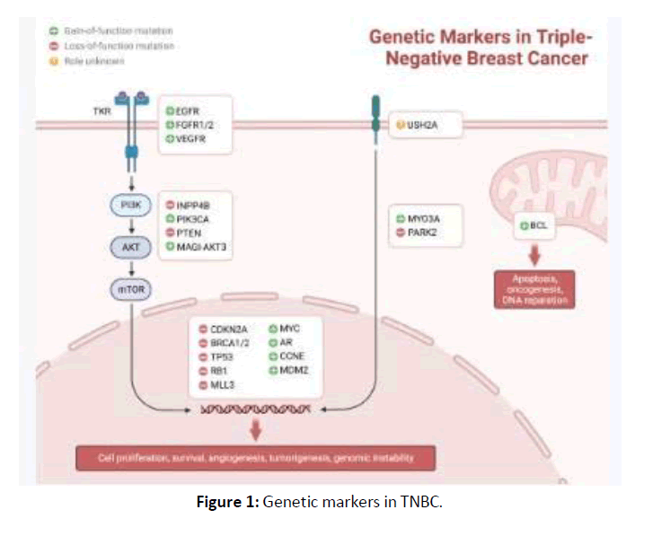
Figure 1. Genetic markers in TNBC.
Clinical Significance of TNBC
TNBC holds a unique concern global wide with advances in clinical outcomes due to aggressiveness poor prognosis associated with the subtype and lack of targeted therapies. TNBC tumours are characterised with high histological grade, high mutation potential, rapid cell division and high metastatic potential compared to other hormone positive breast cancer subtypes. TNBC tumours majorly metastasize to visceral organs like lung, liver, Brain commonly than bone and exhibits early recurrence with in 3-5 years after initial diagnosis.
Poor Prognosis & Lack of Targeted Therapies
Absence of response to hormone and HER2 receptors limit the response to chemotherapy initially and often suffers with resistance and relapse in later stages. TNBC patients usually have low overall survival and disease-free survival compared to patients with hormone receptor–positive or HER2-positive breast cancers. Heterogenicity is possible in TNBC patients responding to therapies differently with variable prognosis. Unlike ER-positive or HER2-positive cancers TNBC lacks the universal effective targeted therapy but progress in terms of medication like PARP inhibitors (for BRCA-mutated TNBC) and immune checkpoint inhibitors (such as pembrolizumab and atezolizumab) occurred recently but these treatments can benefit the patients of few subtypes there by chemotherapy as one of the first line therapy. Thus, there is a urgent need of developing some novel therapies for the treatment of TNBC subtypes universally.
Additional risk stratification
Incidence of Luminal-B breast cancer will be considerably higher in patients with enhancement of parenchymal tissue in organ and the overall statistical difference is significant in all Luminal-B and Luminal-A, HER-2 over-expressed type, and triple-negative type with P value <0.01. The highest percentage of TNBC is associated with patients having marked BPE with statistical significance of P<0.05 between the triple-negative type and the Luminal-A, Luminal-B, and HER-2 over-expression types [Figure 2].
Figure 2. Luminal-A and B Luminal-A breast cancer with ER/PR positive.
Morphology and Pathology of Triple Negative Breast Cancer
Histological Features
70-80% of TNBCS majorly characterised as high-grade invasive ductal carcinomas (IDC) exhibiting the most aggressive biologic nature in tumours.
Grade and Differentiation
According to the Nottingham histological grading system majority of TNBCs belong to grade III with poor differentiation, marked nuclear atypia, and high mitotic activity. Tumour cells often exhibit pleomorphic, vesicular nuclei, prominent nucleoli, and frequent atypical mitoses, there by associated with rapid proliferative capacity. Growth pattern is observed like solid, sheet-like, or nested with less defined well formed glandular structures observed in lower grade breast cancers.
Necrosis and Stromal Features
Necrosis at local area of the tumour is normally observed due to rapid growth associated there by outpacing the blood supply. Stroma often displayed with desmoplastic reaction and as well as dense tumour-infiltrating lymphocytes (TILs). Interestingly high infiltration of lymphocytes (TILs) normally forms a basis for the treatment response to chemotherapy or immunotherapy.
Proliferation Markers
TNBC frequently express high levels of Ki-67, a marker of cellular proliferation, further responsible for their aggressive behaviour in growth and their kinetics.
Morphological Diversity
Invasive ductal carcinoma is recognised as the major dominant histological form TNBC encompasses a group of tumours that differ morphologically contributing to tumour molecular heterogenicity. TNBC phenotype is associated with several types of variants: include [Figure 3]
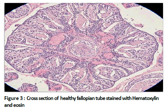
Figure 3. Cross section of healthy fallopian tube stained with Hematoxylin and eosin.
Medullary Carcinoma and Medullary-Like Features
Although Classic medullary carcinoma, is usually rare it is often represented as one of the subtypes of TNBC. >75% of tumour cells exhibit syncytial growth pattern histologically, high nuclear grade, prominent nucleoli, and a dense lymphoplasmacytic infiltrate. Despite its high histological grade MC shows high prognosis compared to TNBC.
Apocrine Carcinoma
Some of the subtypes of TNBC often exhibit apocrine differentiation characterized by large polygonal cells with abundant eosinophilic, granular cytoplasm and prominent nucleoli. These tumours come under Luminal Androgen receptor subtype and respond well with Androgen receptor (AR) targeted therapies.
Metaplastic Carcinoma and its sub types:
Metaplastic carcinomas are usually rare but usually characterised with triple negative phenotype, epithelial-mesenchymal transition and histological heterogenicity.
• Squamous cell carcinoma
• Spindle cell carcinoma
• Chondroid or osseous differentiation (carcinoma with heterologous elements)
• Metaplastic carcinomas tend to possess low prognosis due to insignificant response to chemotherapy.
Diagnosis of TNBC using Immunohistochemistry:
Accurate diagnosis of TNBC depends on diagnosis by immunohistochemistry and is essential for finding the positivity for ER, HER2 and PR and for identification of specific characteristics in tumour. Tumours with <1% nuclear staining for ER or PR identified by immunohistochemistry is classified as negative according to ASCO/CAP guidelines.
HER2 positivity is assessed initially by using IHC and tumours with equivocal 2+ grading is further characterised using fluorescent in situ hybridisation (FISH) to evaluate gene amplification. TNBC are usually defined with HER2 negativity (0 or 1+ IHC, or non-amplified by FISH) and possess basal like phenotype by expressing key markers like basal cytokeratin (CK5/6, CK14, CK17) and epidermal growth factor receptor (EGFR). These markers are may not be important diagnostically but possess high prognosis and response.
Ki-67 expression is highly constituent along with the rapid proliferative capacity in TNBC and Additional markers such as p53 (often mutated and overexpressed) are utilised in research or to know the prognosis of the disease. IHC can help in distinguishing other triple negatives from TNBC such as with distinct subtypes such as lymphomas, sarcomas, and metastatic carcinomas to the breast.
TNBC are often diagnosed with aggressive phenotype compared to other triple negative cancers and due to rapid growth rate and higher histological grade associated with the phenotype patients can be diagnosed with the symptoms earlier before the disease can progress to advanced stages.
Symptoms of TNBC:
The most common feature observed in TNBC is a palpable breast lump and TNBC tumours are usually firm, irregular, and non-tender on examination and tend to be large compared to other hormone positive cancers due to rapid proliferation in the breast tissue. Although most of the TNBC are painless some patients reported with local pain and discomfort due to tissue enlargement by rapid cell division or due to invasion to surrounding tissues. TNBC is characterized by high mitotic index and short doubling time finally causing tissue enlargement in over week to months considerably. This aggressive feature distinguishes TNBC from slow-growing ER+ cancers.
Skin retraction, dimpling, erythema, or thickening may occur in initial stages due to invading of superficial tissues. Progression to advance stages may cause inflammation like redness in the localised tissue. Due to early lymphatic spread, patients may suffer with features like palpable axillary lymphadenopathy. Weight loss, fatigue, bone pain, or neurological symptoms commonly observed in metastatic disease. Diagnosis of TNBC is possible by combinatorial techniques like clinical assessment, imaging, histopathology, and molecular profiling. Early and timely accurate diagnosis is required to plan for effective therapeutic treatment in TNBC.
Imaging Studies for Tumour detection in TNBC:
Imaging helps in early tumour detection, characterization, staging, and surgical planning. Imaging remains first line tool for breast cancer detection. TNBC tumours appear as high-density masses with irregular or spiculated margins through imaging and microcalcifications is less common when compared with ER+ cancers making detection as a most challenging task in few cases. In young patients it is difficult to diagnose breast tumours by mammography due to lack of sensitivity and can be diagnosed using ultra sound imaging with typical features like: include
• Hypoechoic appearance (darker than surrounding tissue).
• Irregular, circumscribed, or micro lobulated margins.
• Posterior acoustic enhancement, a common finding in highly cellular tumours.
• Also helps evaluate axillary lymph nodes.
Magnetic Resonance Imaging (MRI)
MRI can be used to detect highly sensitive multifocal, multicentric, and contralateral breast cancers. TNBC lesions typically show rim enhancement and consistent with high vascularity and aggressiveness. MRI is proven to be valuable for planning surgery and to treat the patients by neoadjuvant chemotherapy.
Cancer studies using Histopathology techniques:
1. Core Needle Biopsy (CNB)
It is the gold standard method used for tissue diagnosis by histology, immunohistochemistry (IHC), and molecular studies. Fine needle aspiration method is not widely accepted due to limited tissue availability.
2. Immunohistochemistry (IHC) Testing
This method is useful for tissue grade classification in TNBC. Tumour samples were tested for Estrogen receptor (ER) Progesterone receptor (PR) & HER2 protein overexpression and/or gene amplification and TNBC diagnosed with negative for all the three receptors and Ki-67 proliferation is usually associated with rapid proliferation and growth rate.
Histological Features
Most of TNBCs are recognised as high grade invasive ductal carcinomas (IDC-NOS) with high mitotic division, necrosis and pushing borders. Histological subtypes of TNBC especially include medullary, apocrine, and metaplastic variants. Due to molecular heterogenicity associated with TNBC high level techniques of molecular profiling is applied for the characterisation of the tumour tissue.
Genetic Testing
Tests related to BRCA1/2 mutation recommended, as BRCA-related TNBCs may get advantage of treatment possible by the use of either PARP inhibitors or chemotherapy. Multigene testing of PALB2, TP53, RAD51, PTEN, and others should be considered for diagnosis and treatment.
1. Molecular Subtyping: Based on the type of gene expression, TNBC can be subclassified into basal-like, immunomodulatory, mesenchymal, luminal androgen receptor (LAR), and others This classification can help to guide towards clinical trials and personalised treatment therapy
2. PD-L1 Expression Testing: This testing is usually performed in cases with advanced and metastatic TNBC and determines possibility of treatment using immune checkpoint inhibitors (e.g., pembrolizumab, atezolizumab).
3. Next-Generation Sequencing (NGS): NGS can help in diagnosis of advanced metastatic tumours and provide detailed information about tumour mutational burden (TMB), homologous recombination deficiency (HRD) and other related mutations.
Current Treatment Strategies
Conventional therapies
Surgery
Surgical management remains the cornerstone for the treatment of localized TNBC and opting for surgery in TNBC depends on tumour size, location, stage, and patient preference.
Lumpectomy (Breast-Conserving Surgery)
Lumpectomy involves removal of tumour with surrounding healthy margin of tissue followed by radiation therapy to avoid /reduce remittance or relapse and usually preferred in early-stage disease
Mastectomy
Mastectomy is a type of surgery which involves removal of entire breast and recommended in the patients diagnosed with features like
• Large or multifocal tumours.
• Contraindications to radiation therapy.
• BRCA mutation carriers
Axillary Management
Sentinel lymph node biopsy (SLNB): standard for node-negative patients.
Axillary lymph node dissection (ALND): performed when diagnosed with nodes positive
Invasive approaches are used minimally to reduce the complications and surgery alone can be rarely sufficient in TNBC due to high risk of systemic relapse, necessitating chemotherapy or other systemic treatments.
Chemotherapy
Chemotherapy is the preferred systemic treatment for TNBC and used as apart of neoadjuvant, adjuvant and metastatic stages.
Neoadjuvant Chemotherapy (NAC)
Neoadjuvant chemotherapy is the preferred treatment before to surgery to mainly shrink the tumours allowing for breast conserving surgery. Pathological complete response in TNBC is about 30-50% than other subtypes and a predictive of better long-term outcomes. Standard regimens are possible with medical agents like anthracyclines + taxanes and with help of platinum agents.
Adjuvant Chemotherapy
Adjuvant chemotherapy is preferred in patients with the risk of recurrence after surgery. Similar regimens are followed in adjuvant chemotherapy as that of neoadjuvant chemotherapy.
Chemotherapy Agents Commonly Used in TNBC
Anthracyclines (doxorubicin, epirubicin): These belongs to class of DNA intercalators that induce apoptosis. Taxanes (paclitaxel, docetaxel): These drugs inhibit microtubule depolymerization, there by inhibiting cell division.
Platinum agents (cisplatin, carboplatin): Highly effective in the patients with BRCA1/2 mutations as these tumours are deficit with DNA repair mechanisms.
Limitations of Chemotherapy Chemoresistance:
In many of the TNBC patients breast cancer usually relapse after initial response associated with Cardiotoxicity, neuropathy, nephrotoxicity/ototoxicity. The patients usually suffer with early relapse risk typically with in 3-5 years of initial diagnosis.
Radiation Therapy
Radiation therapy plays vital role in treatment of cancer disease control after surgery.
Indications:
• Patients undergoing with lumpectomy (mandatory in breast-conserving therapy).
• In patients treated with mastectomy and after mastectomy for tumours >5 cm or node-positive disease.
• As a treatment for patients possessing bone, brain, or chest wall metastases.
Benefits:
• Positive outcome with significant reduction in relapse rates
• Increases overall survival rate in TNBC patients with few of the subtypes.
Targeted and Novel Therapies
Limitations of conventional therapy had led to development of targeted therapies at the molecular level and immunotherapy- targeted therapy some of which are already applicable and effective in clinical use.
PARP Inhibitors
Usage of PARP inhibitors gained importance with patients those are particularly possessing BRCA-mutant or homologous recombination–deficient (HRD) TNBC. PARP enzymes are involved in repair of ssDNA breaks inhibition by inhibitors there by causing accumulation of DNA damage and lethal in cells with BRCA mutations or BRCA-/-.Approved drugs as PARP inhibitors include Olaparib (OlympiAD trial), Talazoparib (EMBRACA trial). Clinical benefits include Improved progression-free survival (PFS) in metastatic BRCA- mutant TNBC compared to chemotherapy and less toxic regimens are possible compared to standard cytotoxic regimens.
Immune Checkpoint Inhibitors
TNBC is associated with heightened immunogenic response compared to other breast cancers because of higher levels of infiltrating lymphocytes (TILs), PD-L1 expression, and tumour mutational burden.
PD-1/PD-L1 Inhibitors:
Pembrolizumab (anti–PD-1):
Approved for combinatorial chemotherapy for the treatment of PD-L1–positive metastatic TNBC. And high-risk early TNBC as part of neoadjuvant/adjuvant therapy.
Atezolizumab (anti–PD-L1): Used as treatment in combination with nab-paclitaxel in PD-L1–positive advanced TNBC.
Clinical Benefits include prolonged progression-free survival and overall survival in advanced TNBCs. Positive response is seen in cases with PD-L1 positive and immune-inflamed tumours.
Antibody-Drug Conjugates (ADCs)
ADCs belongs to new class of drugs that allow targeted killing of cancer cells with monoclonal antibody linked to toxin.
Sacituzumab Govitecan (Trop-2–directed ADC):
This drug is mainly recommended in case of pretreated metastatic TNBC and the drug includes SN-38 as the cytotoxic drug particle. Sacituzumab Govitecan is proven with significant improvement in survival compared to standard chemotherapy.
Emerging ADCs:
Ladiratuzumab Vedotin mainly targets LIV-1 and HER2-low ADCs are under study for TNBC patients which show low-level expression of HER2.
Clinical Trials and Ongoing Research
Most of the clinical trials and ongoing research suggest the improvement in targeted therapies in the treatment of TNBC. These majorly include:
Combination Therapies
ICIs + chemotherapy.
PARP inhibitors + ICIs (synergistic effect via DNA damage–induced neoantigens).
ADCs + checkpoint inhibitors.
Targeted Small Molecule Inhibitors
PI3K/AKT/mTOR inhibitors in TNBC in case of mutated signalling agents of the pathway
Androgen receptor (AR) inhibitors for the treatment of luminal AR subtype cancers.
Immunotherapy Advances
Cancer vaccines targeting tumour cells and tissues by activating cellular response
CAR-T and CAR-NK therapies targeting EGFR, Trop-2, and PD-L1.
Precision Oncology Approaches
Liquid biopsies (ctDNA, CTCs) for relapse detection.
Next-generation sequencing (NGS) for cancer treatment which is particularly biomarker driven
Prognosis and Challenges
High relapse rates (within 3–5 years). Metastasis (lung, brain, liver). Restriction of treatment to ER+/HER2+ cancers and lack of therapeutic effect in other than ER+/HER2+ cancers.
References
- Bianchini G, Balko JM, Mayer IA, Sanders ME, Gianni L. Triple-negative breast cancer: challenges and opportunities of a heterogeneous disease. Nature Reviews Clinical Oncology. 2016;13:674-690.
- Lehmann BD, Pietenpol JA. Identification and use of biomarkers in treatment strategies for triple-negative breast cancer subtypes. Journal of Pathology. 2014;232:142-150.
- Schmid P, Adams S, Rugo HS, Schneeweiss A, Barrios CH, et al. Atezolizumab and nab-paclitaxel in advanced triple-negative breast cancer. New England Journal of Medicine. 2018;379:2108-2121.
- National Comprehensive Cancer Network. NCCN Clinical Practice Guidelines in Oncology: Breast Cancer. Version 2023. NCCN Guidelines. 2023.
- Early Breast Cancer Trialists' Collaborative Group. Effects of radiotherapy and of differences in the extent of surgery for early breast cancer on local recurrence and 15-year survival. Lancet. 2005;366:2087-2106.
- Cortazar P, Zhang L, Untch M, Mehta K, Costantino JP, et al. Pathological complete response and long-term clinical benefit in breast cancer: the CTNeoBC pooled analysis. Lancet. 2014;384:164-172.
- Schmid P, Cortes J, Pusztai L, McArthur H, Kümmel S, et al. Neoadjuvant chemotherapy in triple-negative breast cancer: current practice and novel approaches. Annals of Oncology. 2018;29:44-56.
- Tutt A, Tovey H, Cheang MCU, Kernaghan S, Kilburn L, et al. Carboplatin in BRCA1/2-mutated and triple-negative breast cancer. Lancet Oncology. 2018;19:475-487.
- McGale P, Taylor C, Correa C, Cutter D, Duane F, et al. Effect of radiotherapy after mastectomy and axillary surgery on 10-year recurrence and 20-year breast cancer mortality. Lancet. 2014;383:2127-2135.
[Cross ref], [Google scholar], [PubMed]
[Cross ref], [Google scholar], [PubMed]
[Cross ref], [Google scholar], [PubMed]
[Cross ref], [Google scholar], [PubMed]
[Cross ref], [Google scholar], [PubMed]
[Cross ref], [Google scholar], [PubMed]
[Cross ref], [Google scholar], [PubMed]